


The high incidence of keratoconus has caused its management, etiology, and pathogenesis to be controversial topics in the ophthalmology field. This study aims to analyze the relationship between the different publications and authors through citation networks, as well as to identify the research areas and determine the most cited article.
MethodsThe search for publications was carried out through the Web of Science database, using the term “Keratoconus” between 1900 and December 2022. The Citation Network Explorer and CiteSpace software were used for the publication analysis.
Results9,655 publications were found, with 124,379 citations generated on the network. The year with the highest number of publications was 2021. The most cited publication was “Keratoconus” by Rabinowitz, published in 1998. Cluster function gave five groups of research areas about keratoconus: corneal signs and parameters, cross-linking efficiency and effects, clinical factors, keratoplasty, and treatment.
ConclusionsThe citation network offers an objective and comprehensive analysis of the papers on keratoconus.
Burchard Mauchart, a German professor, described keratoconus for the first time in 1748 as “Staphyloma Diaphanum”. This condition had been previously discussed by some physicians, who referred to it as “ochlodes” meaning “irritating” in Greek. In 1854, a British doctor, John Nottingham, named it as “chronicle cornea”, and nowadays, many of his ideas are still in use.1
In 1859, William Bowman was the first to use an ophthalmoscope to observe keratoconus. Ten years later, John Horner, a Swiss doctor, introduced the modern name of the affection: keratoconus.2,3
The first lenses fitting to improve keratoconus patients' vision were named “contact lenses” in 1888. From then, contact lenses history, the current name of those lenses, and keratoconus are closely linked.4
The first keratoplasty of this pathology was performed by Anton Elsching in 1930, which led the way to a solution for keratoconus cases with a worse prognosis.5
Nowadays, the medical understanding of keratoconus is extremely sophisticated, and it is differentiated and classified into mild, moderate, and advanced. Although keratoconus can occur in any life stage, younger people (aged between 10 and 25) have a higher risk. The cornea is gradually flattened, and it starts to bulge in keratoconus patients. It often causes high myopia and irregular astigmatism as the disease progresses. The first signs are quick changes in vision that require settings in the patient´s spectacles. Other symptoms are light sensibility, eye fatigue and irritation, halos around lights at night, headaches, and the need to rub their eyes. As it progresses, the corneas acquire a noticeable conical shape.6,7
Keratoconus is the most common dystrophy or degenerative corneal disorder, having an estimated prevalence of approximately 54.5 for 100,000 (0.05 %) and with an annual incidence of 4.6 for 2000 inhabitants.8 Many symptoms of keratoconus are similar to other corneal disorders, especially at the beginning of the affection. This causes keratoconus may be difficult to diagnose.
Nowadays, there is controversy about why some people develop keratoconus. Until recently, it was considered a non-inflammatory disease, but nowadays there is evidence against this assumption.9 Main theories point at genetics, environment, and hormones. Some clinicians have perceived a slightly higher possibility of developing keratoconus in people with a family record of it, but this correlation has not been proved. The environmental causes include allergies, making patients excessively rub their eyes, or wearing wrong-fitted contact lenses. Some researchers hypothesized that keratoconus is related to the endocrine system because its onset often occurs at puberty and worsens during pregnancy.10–12
Generally, the cornea stabilizes, so it can be corrected by wearing spectacles (mild stage) or contact lenses (moderate or advanced stage). However, 10 and 20 percent of keratoconus patients have serious problems due to a lack of stabilization, and they will require alternative treatments, such as cross-linking and intrastromal corneal rings. Regarding the most severe cases, corneal transplantation could be needed.13
Although great advances have been made recently, there is still a lot of controversy and knowledge limitations, especially regarding an early diagnosis, its evolution control, and more efficient treatments. Therefore, present and future investigations are based on these aspects.14
Citation network analysis is used to search scientific literature on a specific subject. In other words, through a single publication, it is possible to find other additional relevant publications to demonstrate, both qualitatively and quantitatively, the relationships that exist between articles and authors.15,16
This study aimed to show a citation network analysis of keratoconus, identifying the different research fields, and authors. It also aimed to determine the most cited publications and the relationship between publications and research groups. To sum up, the main objective of this study is to know the development of scientific literature in the keratoconus research field.
Materials and methodsDatabaseThe search of different publications was carried out using the Web of Science (WOS) database, with the following search term: “Keratoconus”. Web of Science makes it possible to add references to your library when conducting bibliographic searches directly in external databases or library catalogs. The search was carried out by selecting the Subject as the search field, and it was limited by abstract, title, and keywords.
About the citation indexes, the Social Sciences Citation Index, the Science Citation Index Expanded, and the Emerging Sources Citation Index were used.
On the other hand, and given that how some authors and institutions cite works may vary, the CiteSpace software was used to standardize the data.
Data analysisThe publications were analyzed using the Citation Network Explorer software, which allows the researcher to analyze and visualize citation networks of scientific publications. Likewise, it is possible to download citation networks directly from the Web of Science and manage citation networks, including millions of publications and related citations. A citation network of several millions of publications can be the starting point for a deeper analysis to obtain a small subnetwork with 100 publications on the same subject.
A quantitative analysis of the most mentioned publications in a period was carried out using the attribute Citation score. Therefore, not only the internal connections within the Web of Science database were quantified, but also any external connections, meaning that other databases were considered.17
Citnetexplorer provides several techniques for the analysis of publications' citation networks. The clustering functionality is achieved using the formula developed by Van Eck in 2012.17

Then, to assign a group to each publication, the Clustering functionality was applied. As a result, the most related publications are usually found in the same group based on the citation networks.17
Finally, the core publications were analyzed using the Identifying Core Publications functionality, consisting of identifying the publications that are considered the core of a citation network because they are a minimum number of connections with other core publications and eliminating the irrelevant ones. The number of connections is established by the researchers, so the higher the value of this parameter is, the lower the number of core publications.17 In this study, the publications with at least four citations in the citation network were considered.
On the other hand, the drilling down functionality allows for a deeper analysis of each group at different levels.
The CiteSpace (5.6.R2) software was used to conduct the scientometric analysis. This software, developed by Chen Chaomei, is Java-based and it is comprised of five basic theoretical aspects: Kuhn´s model of scientific revolutions, Price's scientific frontier theory, the organization of ideas, the best information foraging theory of scientific communication, and the theory of discrete and reorganized knowledge units.18,19 In the scientometric analysis process, there are also some parameter indicators to carry out a specific assessment. The H-Index is a mixed quantitative index, suggested by George Hirsch from the University of California, United States. It evaluates the quantity and level of academic output of researchers and institutions. The H-Index indicates that h out of N published articles in a journal have been cited at least h times.20 The Degree indicates the number of connections among the authors (organizations, countries) in the co-occurrence knowledge graph. A higher value in this Degree indicates a greater level of communication and collaboration between the authors (organizations, countries). Besides, the centrality value measures the importance of the nodes within the research cooperation network, and the half-life is a parameter that represents the continuity of institutional research from a time perspective.18
ResultsThe first articles on keratoconus were published at the beginning of 1900, therefore the selected period of study was from 1900 to December 2022. Following the WoS search, 9655 publications and 124,379 citation networks were found. As shown in Fig. 1, the number of publications on keratoconus has increased exponentially since 2008 (1900–2007: 22.8 %; 2008–2022:77.2 %). The year with the highest number of publications was 2021, with 701 publications and 234 citation networks.
Publication descriptions: language, countries, and research areas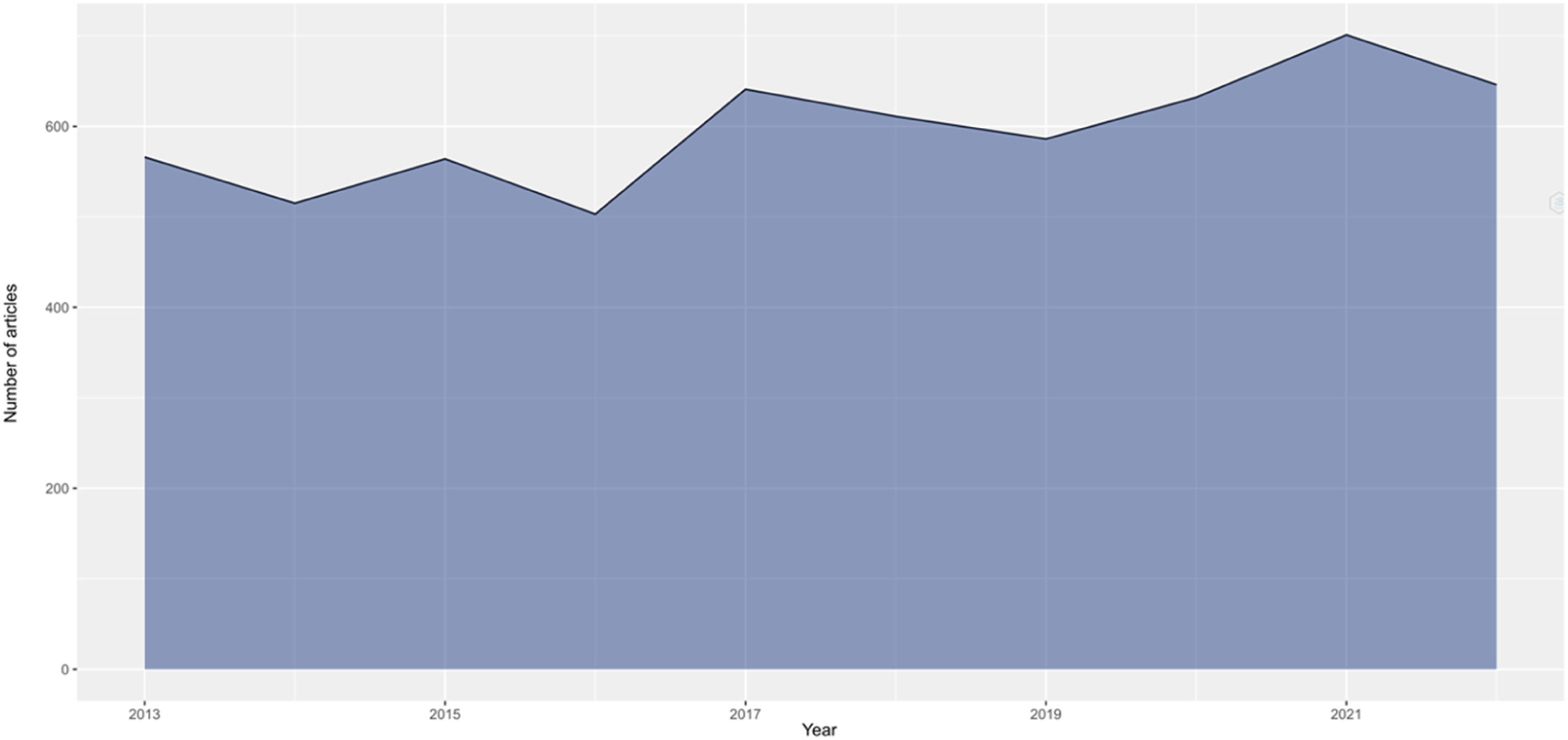
Of all publications, 73.7 % were articles, 11.4 % were abstracts of congresses and conferences, 6.0 % were reviews, 4.9 % were letters to the editor, and 3.6 % were proceeding papers.
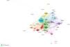
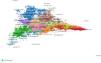
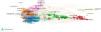
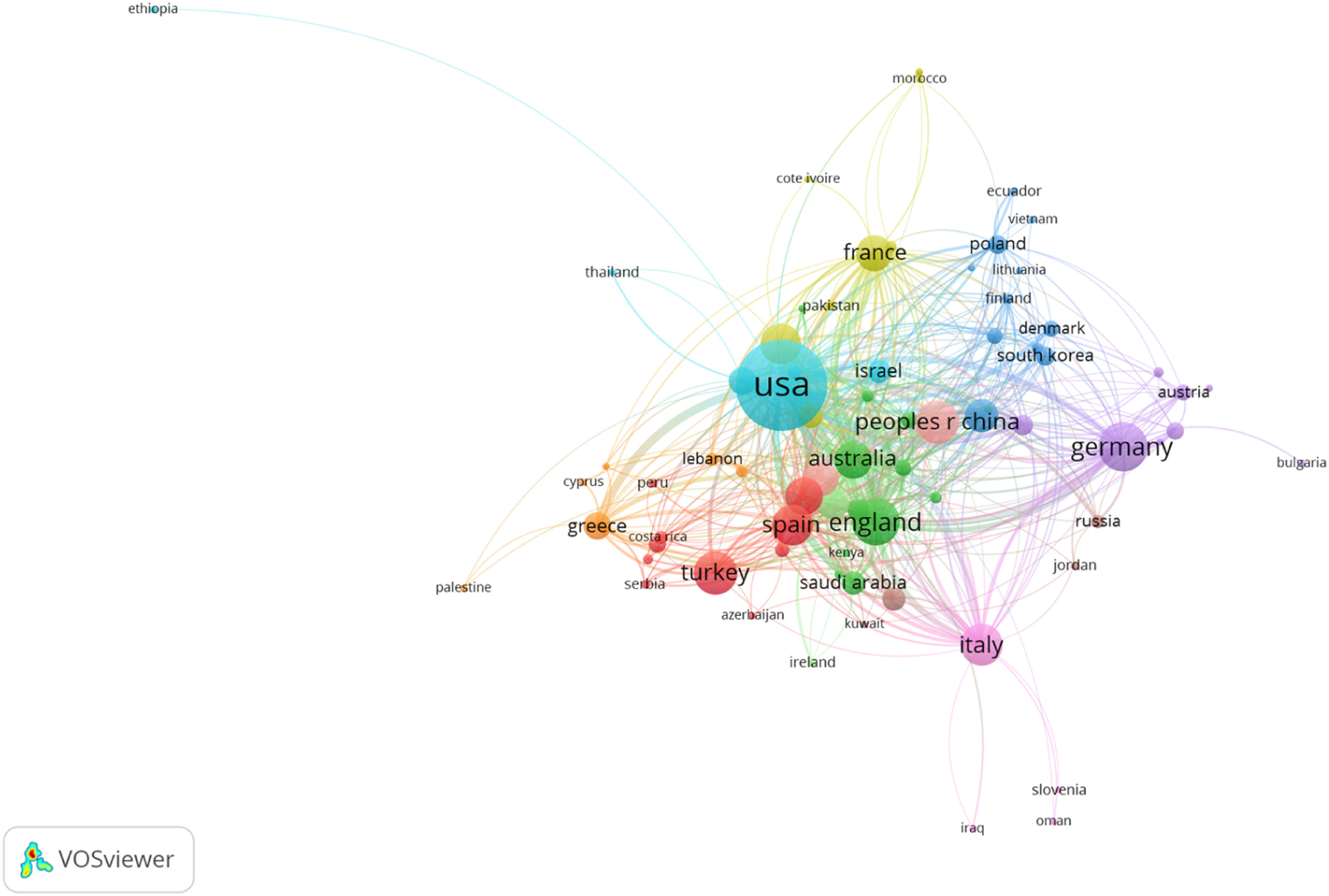
About the language of the publication, 95.1 % were in English, 2.8 % in German and 1.2 % in French. Fig. 2 and Table 1 show the countries with the most publications: the United States (25.0 %), Germany (7.3 %), and England (7.3 %). Fig. 2 shows the most important countries and the group to which they belong. An article's color represents the group, and the lines between the elements represent links. Table 1 shows the main characteristics of the four top groups shown in Fig. 2. Research on this topic is multidisciplinary, but the fields of ophthalmology (84.5 %) and surgery (12.2 %) (Table 2) highlight.
The top 10 research areas.
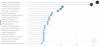
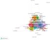
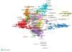
Fig. 3 shows the authors with the highest number of publications on keratoconus: Seitz B (1.8 %), Langenbucher A (1.3 %), and Ambrosio R (1.2 %). Fig. 4 shows the most productive institutions: the Tehran University of Medical Sciences, The Ohio State University, and the Federal University of São Paulo.
Journals, keywords, and most cited publications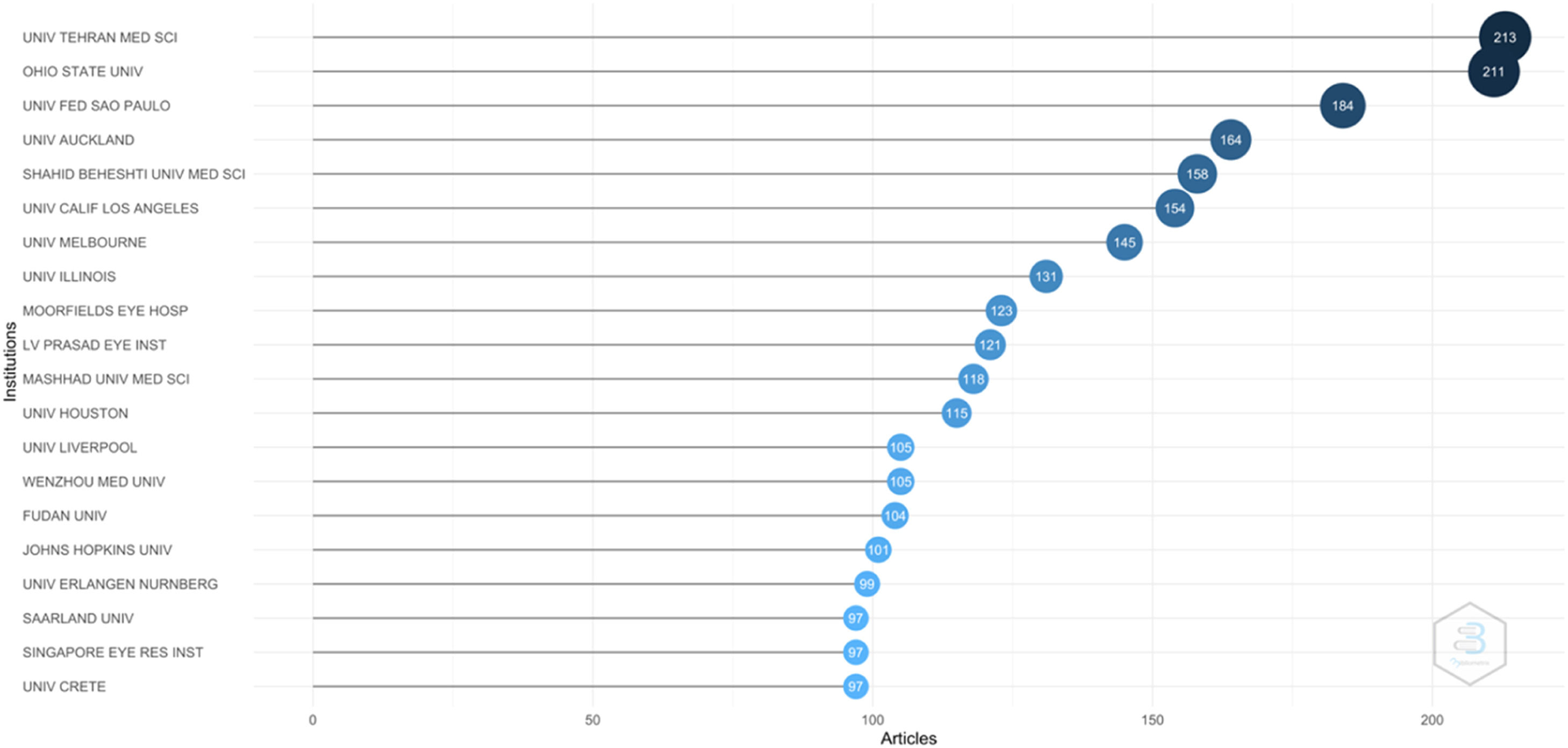
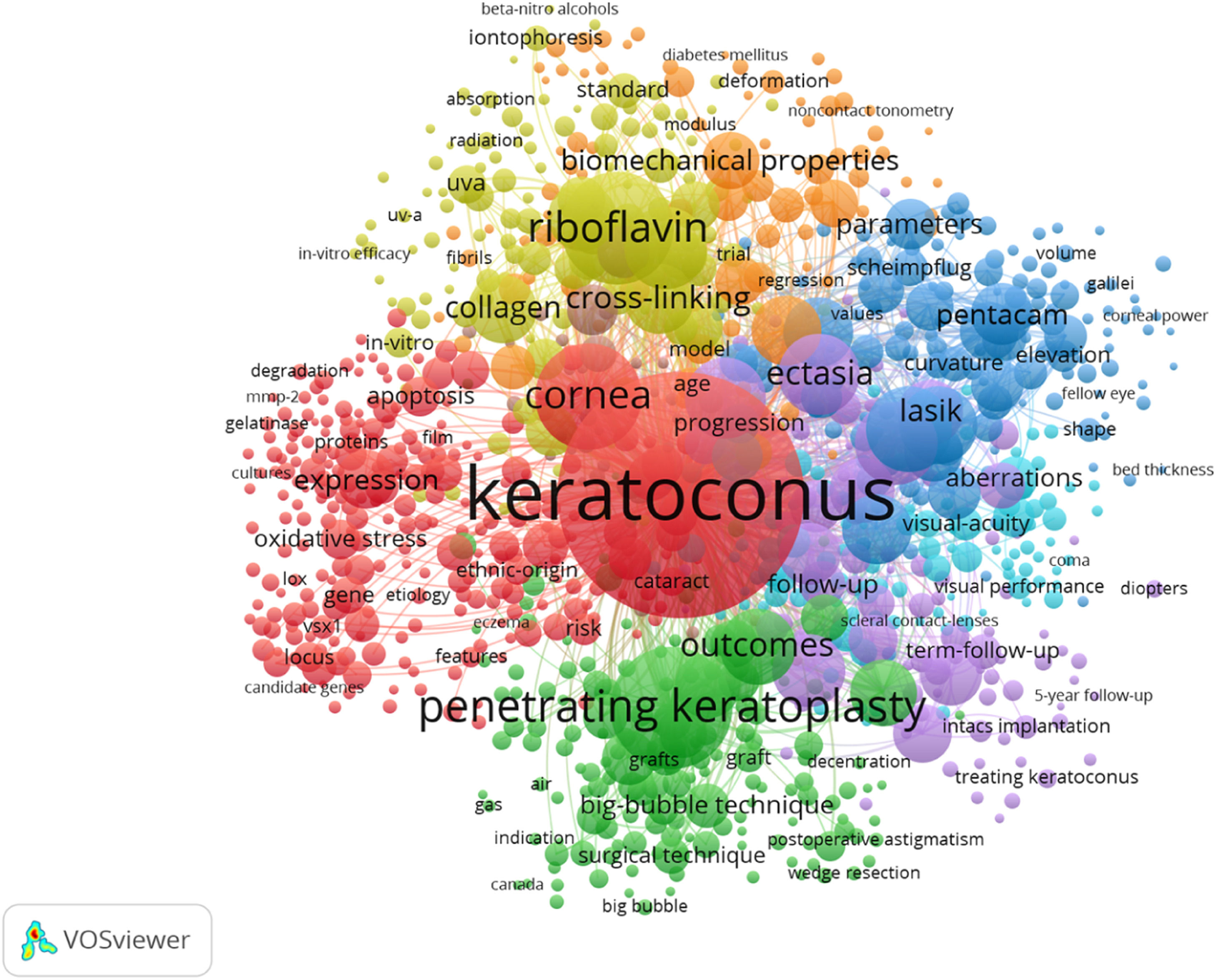
Table 3 shows the main journals that have published about keratoconus and the number of publications according to the WoS database.
The top 10 journals.
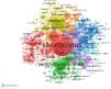
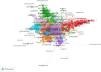
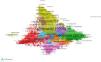
On the other hand, the most used keywords were “Keratoconus” “Penetrating keratoplasty” and “Cornea”. Table 4 and Fig. 5 show the most used keywords in the most relevant publications. Table 5 shows the main characteristics of the top five groups in Fig. 5.
The 20 most used keywords.
Characteristics of the most used keywords.
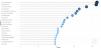
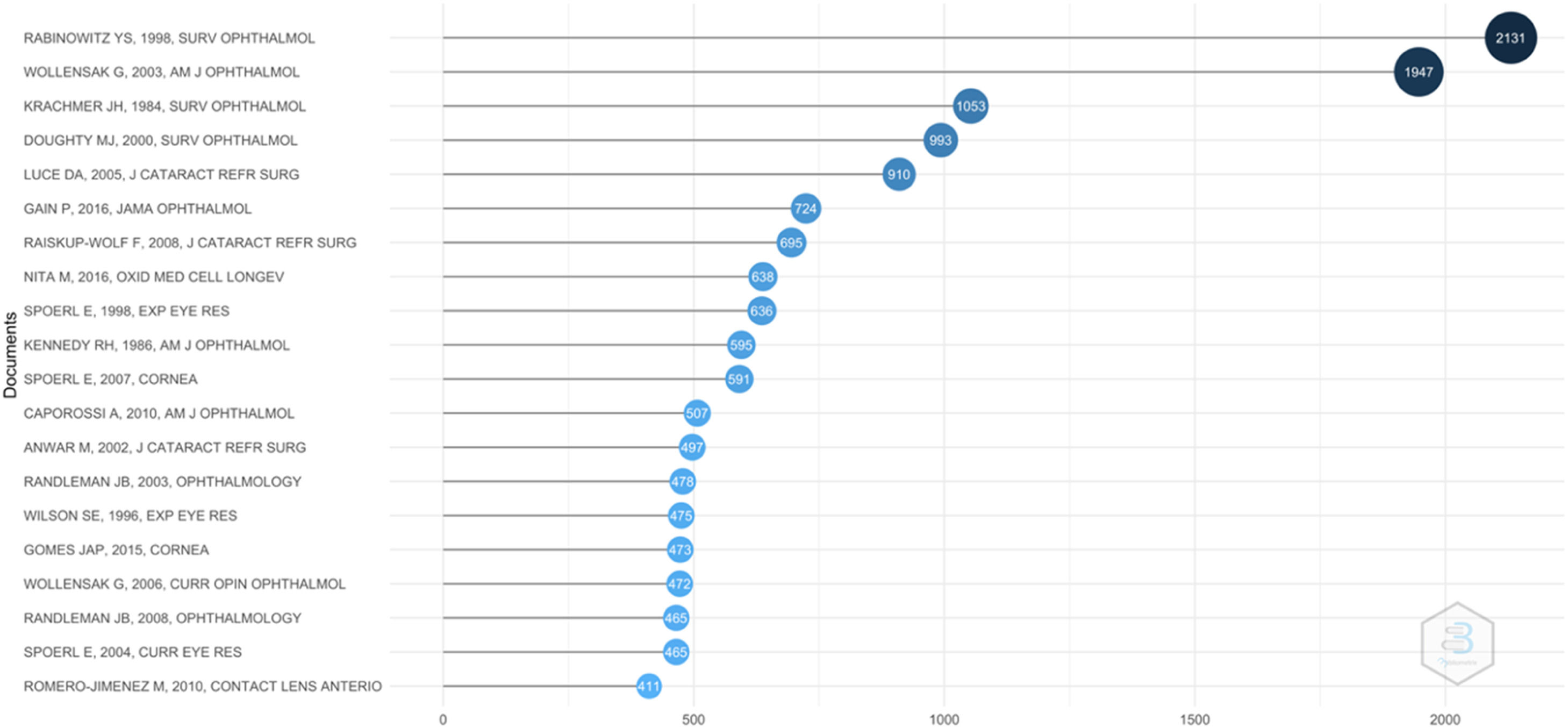
Fig. 6 shows the top 20 articles. The most cited publication was the article by Rabinowitz, published in 1998 with a citation index of 486.
When analyzing the top 20 articles, four of them treat the importance of clinical factors associated with keratoconus, 14 of them the efficiency and effects of cross-linking, and the other two, the corneal signs and parameters, and the keratoconus treatment with corneal ring segments.
Clustering and core functionSix groups were found using the clustering function, of which five had a high number of publications. However, the remaining group only represents 0.44 % of them.
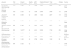
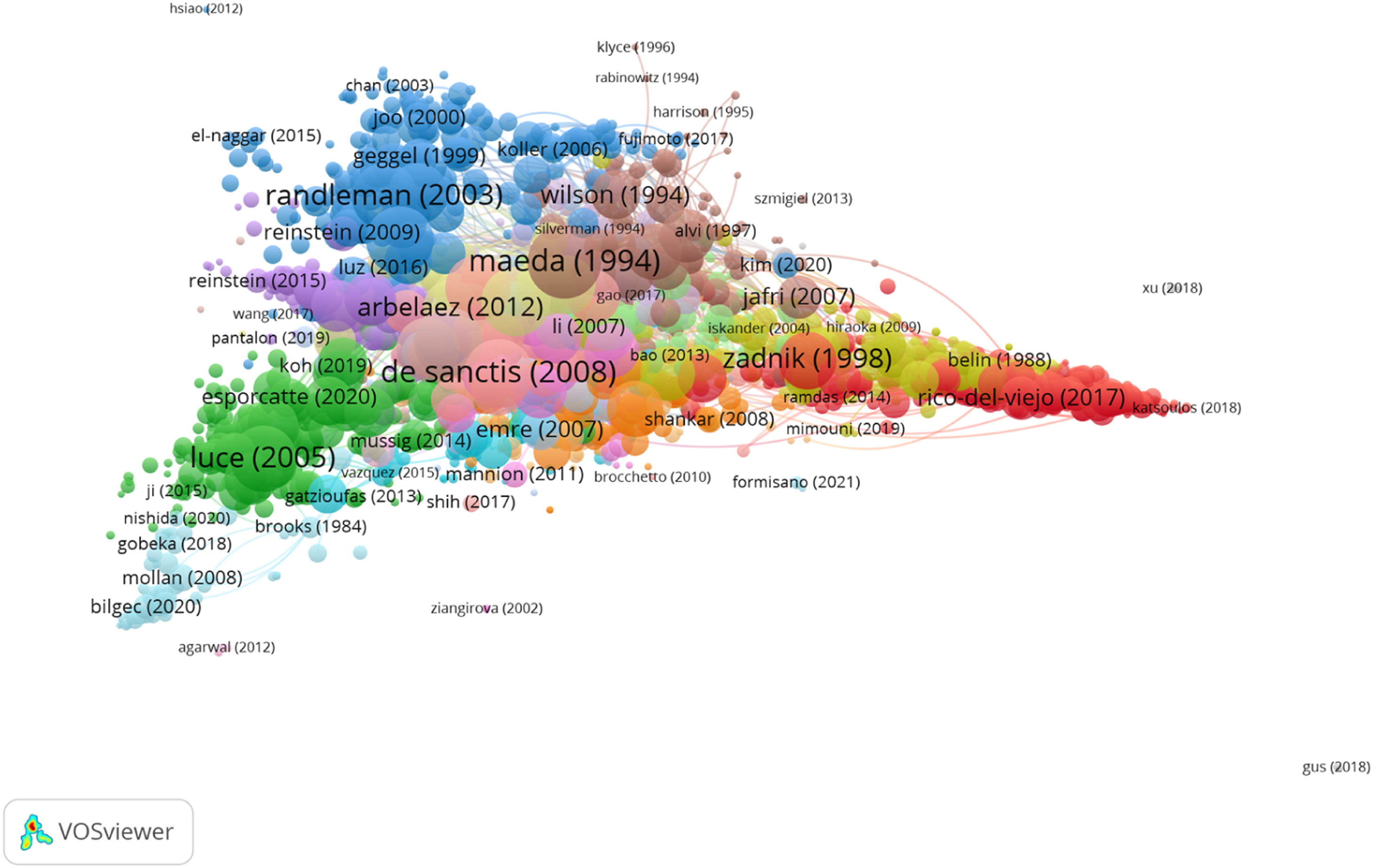
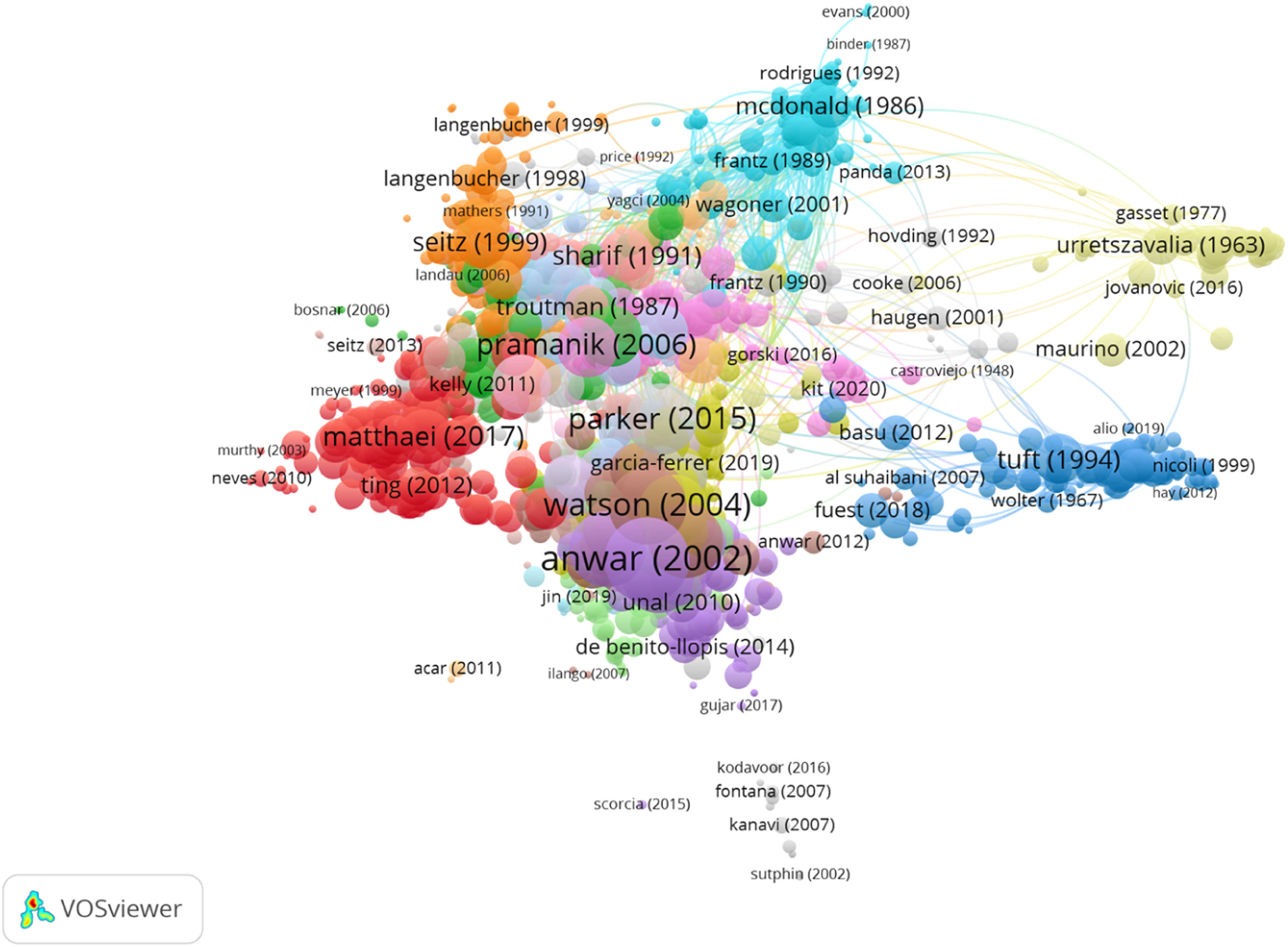
Group 1 is composed of 1785 publications and 18,094 citations. The most cited publication was the article by Zadnik et al.,21 published in 1998 in the Investigative Ophthalmology & Visual Science. The papers in this group analyzed the corneal signs and parameters in keratoconus patients (Fig. 7).
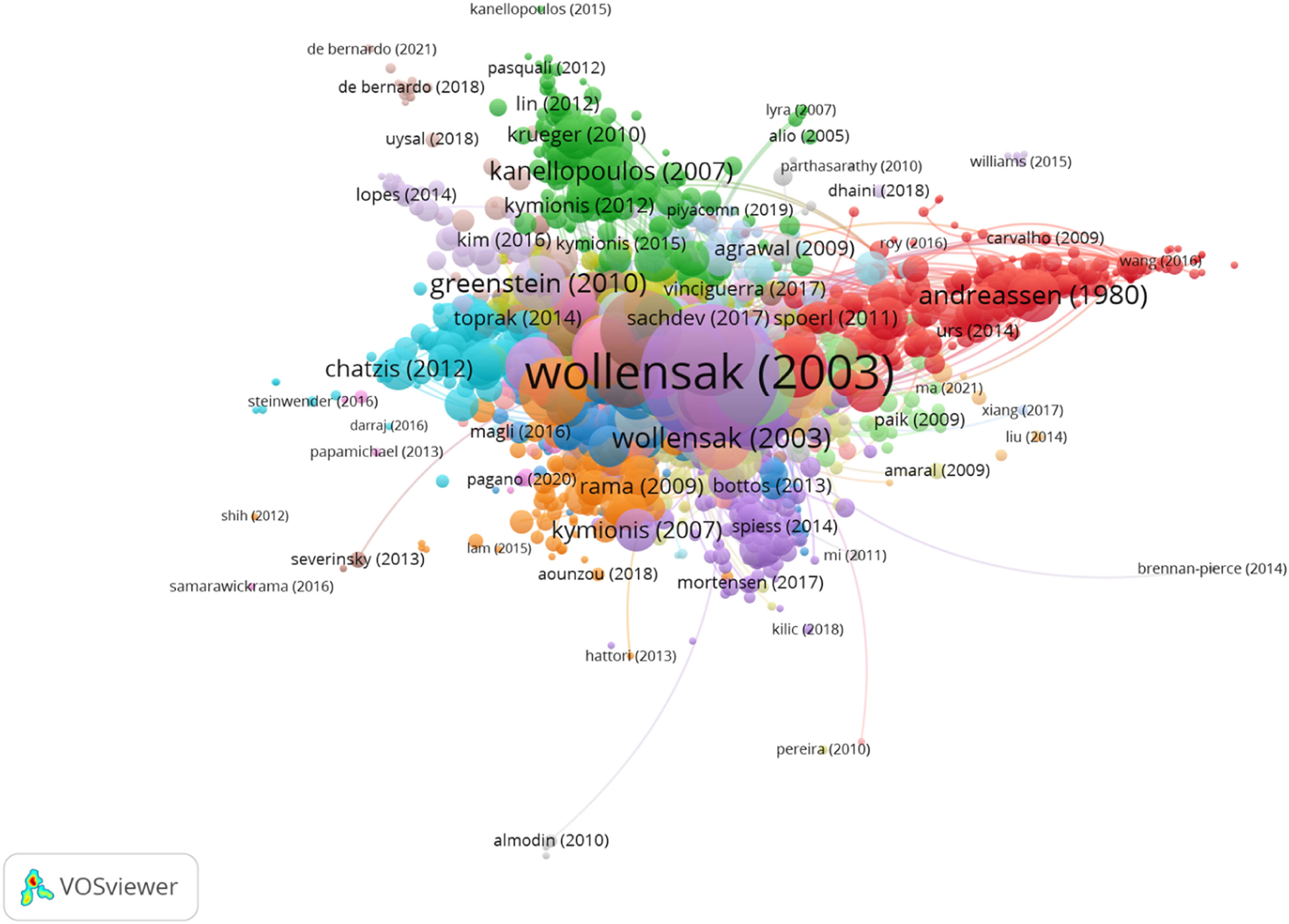
Group 2 consisted of 1730 publications and 27,022 citations. The most cited publication was the article by Wollensak et al.,23 published in 2003 in the American Journal of Ophthalmology. The papers in this group analyzed the efficiency and the effects of crosslinking in keratoconus patients (Fig. 8).
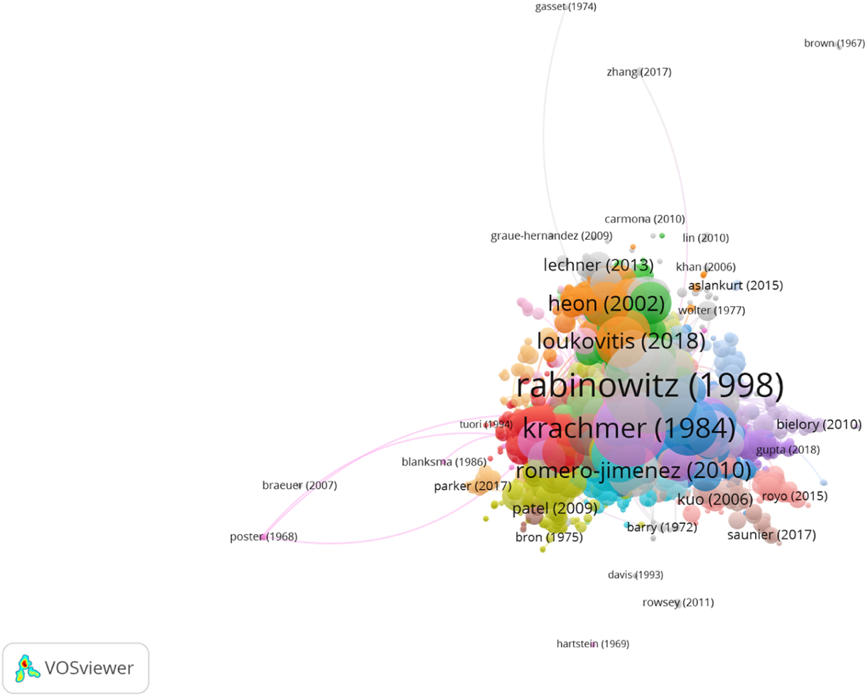
Group 3 was made up of 1707 publications and 18,419 citations. The most cited publication was the article by Rabinowitz 22 published in 1998 in the Survey of Ophthalmology, which is the first most cited publication among a group of 20. The papers in this group analyzed the importance of clinical factors associated with keratoconus (Fig. 9).
Group 4 consisted of 1362 publications and 11,503 citations. The most cited publication was the article by Anwar et al.24 published in 2020 in the Journal of Cataract and Refractive Surgery. The papers in this group analyzed the different techniques of keratoplasty (Fig. 10).
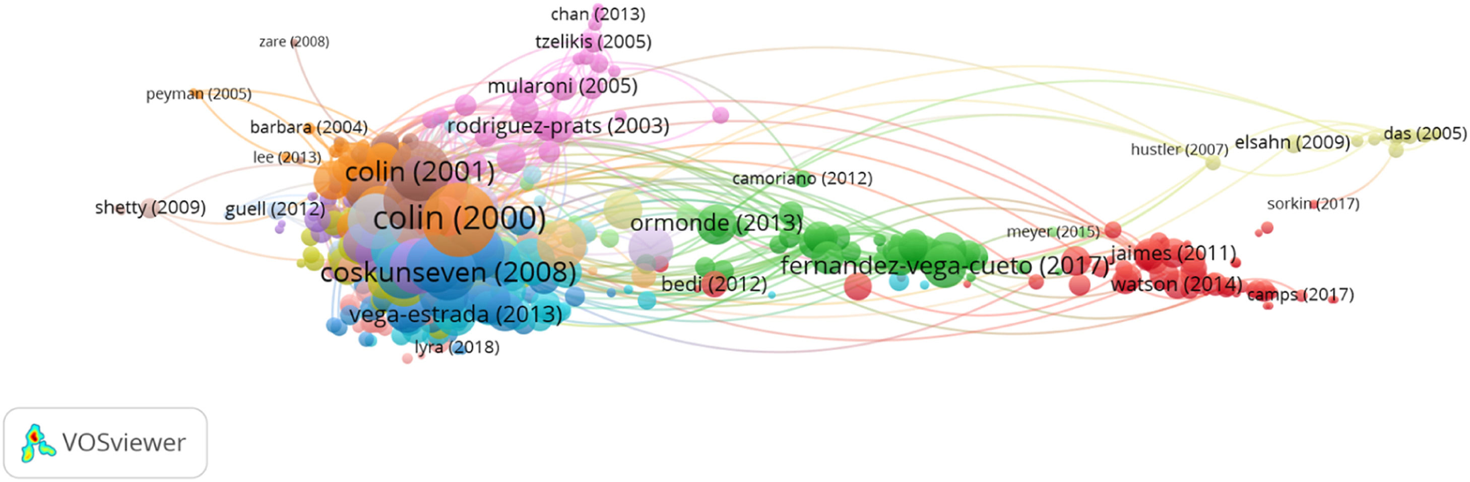
Group 5 was made up of 539 publications and 5366 citations. The most cited publication was the article by Colin et al. 26 published in 2000 in the Journal of Cataract and Refractive Surgery. The papers in this group analyzed keratoconus treatment with intrastromal corneal rings (Fig. 11).

Regarding core function, 7534 publications with at least four citations were found, and the citations network was comprised of 122,781 publications, representing 78.03 %. Moreover, there is a clear approach within the research field as the main discussed subject is related to the corneal signs and parameters in keratoconus patients.
DiscussionThe main databases, such as the WOS or Scopus, allow for the creation of citation networks. However, when conducting a systematic review of all of the existing literature on a subject, their usefulness is limited, given that they do not provide a general overview of the connection between citations of a group of publications. Therefore, the CitNetExplorer software was used to visualize, analyze, and explore the citation networks of scientific publications.17
This study aimed to analyze the existing literature on keratoconus through the WOS database, using the CitNetExplorer software to collect and analyze every available piece of literature on keratoconus to date. Through the analysis of citation networks, it was possible to obtain the connection between the fields of study and the different research groups. The clustering function groups publications according to the relationships that exist among citations, while the drilling down function makes a more in-depth analysis of the bibliography for each group. The core publications function shows the main publications, meaning those with a minimum number of citations. All these functions help to make a complete analysis and study of the research on the field of study.
Nearly 95 % of the publications are in English, with the top three producing countries being the USA, Germany, and England. The case of Germany can be explained because corneal cross-linking was developed at the University of Dresden in 1997.25
The main areas of research are ophthalmology and surgery, which is the reason why the most common keywords are keratoconus, penetrating keratoplasty, and cornea. Moreover, the journals that have a higher number of publications on the subject are Investigative Ophthalmology and Vision Science, Cornea, and the Journal of Cataract and Refractive Surgery.
The three most productive authors are Alió, Shetty, and Hafezi. All of them have contributed with remarkable findings on keratoconus.
Dr. Alió is the author of one of the classifications of keratoconus. By analyzing topography, corrected distance visual acuity (CDVA), K, internal astigmatism (D), coma-like RMS (µm), and corneal asphericity Q at 8 mm.; he classifies keratoconus into 5°: I, II, III, IV, and IV-Plus. In 2016, he published a book entirely focused on the subject: Keratoconus. Recent advances in diagnosis and treatment.27,28
The paper carried out by Dr. Shetty includes the influence of stromal molecular markers on corneal ectasia and risk-scoring systems to predict ectasia after refractive surgery. At the 2015 All India Ophthalmological Society annual conference, Dr. Rohit was awarded the prestigious Col. Rangachari award for the best article at the conference for his work on “Is inflammation driving keratoconus? A Holistic Study of The Molecular Pathways”. 29
Farhad Hafezi was first recognized as a leading retina researcher in 1994, after being the first to discover a gene responsible for some retinal degeneration. However, he changed his research focus to the cornea in 2003, and it is because of this particular work on corneal cross-linking (CXL) that he is known internationally.30
About the institutions, the Federal University of Sao Paulo (Universidade Federal de Sao Paulo) is one of the most important due to the contribution of Paulo Ferrara to the development of one of the most used intrastromal rings (Ferrara rings).31
Regarding the Teheran University of Medical Sciences, the Department of Epidemiology stands out, particularly Doctor Hashemi, who published in 2020 a systematic review and meta-analysis on the prevalence and risk factors of keratoconus.32
Thirdly, The Ohio State University carried out the Collaborative Longitudinal Evaluation of Keratoconus (CLEK) study. The CLEK study was a multicenter study of 1209 patients with keratoconus who were examined annually for eight years. The aims were to prospectively define changes in vision, corneal curvature, corneal status, and vision-specific quality of life in patients with keratoconus.33 The study led to another classification of keratoconus: the Keratoconus Severity Score (KSS).34
So far, the most cited article has been the one by Rabinowitz,22 describing keratoconus as a non-inflammatory ectasia with an incidence of around 1 in 2000 in the general population.
By analyzing the most cited articles, the main subjects are the importance of associated clinical factors, corneal signs and parameters, the efficacy and effects of cross-linking, and the treatment with keratoplasty or intrastromal rings. All these aspects are validated in the clusters that were found.
The corneal signs and parameters are analyzed in patients with keratoconus in group 1. The most cited article of this group analyses the clinical signs of 1209 patients with a mild to moderate degree of keratoconus.21 The findings suggest that keratoconus is not related to further connective tissue disruption. Nevertheless, it is recommended to conduct a study of at least three years and evaluate its impact on life quality.
Group 2 highlights the analysis of the effectiveness and effects of cross-linking. The effectiveness of crosslinking with riboflavin and ultraviolet light to halt the progression of keratoconus is analyzed in the most cited study.23 To this effect, 23 eyes with moderate and advanced keratoconus were analyzed. After central corneal abrasion, riboflavin photosensitizing drops were applied, and the eyes were exposed to UVA (370 nm, 3 mW / cm (2)) at a distance of 1 cm for 30 min. The check-ups were carried out every six months, and the study lasted from three months to 4 years. The results showed that crosslinking can help stop its progression, thus reducing the need for keratoplasty.
Group 3 is made up of articles analyzing the importance of associated clinical factors. The most cited paper is the article carried out by Rabinowitz, as mentioned above.
Group 4 includes articles that analyze various keratoplasty techniques. The most cited article describes the lamellar keratoplasty technique, which consists of inserting air with a disposable needle, deeply and beveled downwards, into the paracentral corneal stroma to detach the central part of the Descemet membrane.24 Then, a small opening is made in the air bubble, and the remaining stromal layers are lifted with an iris spatula, cut with a blade, and removed with scissors.
Finally, group 5 includes articles analyzing the keratoconus treatment with intrastromal rings. The results of the corneal intrastromal rings to correct keratoconus without a central corneal scar are discussed in the most cited article.26 To this effect, various prospective, non-comparative, and interventionist cases are compared, in which Intacs were implanted in 10 eyes with keratoconus and with clear central corneas and contact lens intolerance. It was done after having checked their corneal pachymetry. The results showed that intrastromal rings may reduce corneal protrusion and the astigmatism associated with keratoconus.
Therefore, the number of studies on keratoconus has been increasing, as more research is needed to improve its diagnosis, management, and treatment.
About citation networks, the studies on the subject have also been increasing, as this is the only method of analysis, providing a global overview of the different fields within a specific topic. Moreover, the CitNetExplorer software allows for the analysis of all existing studies on a particular topic, allowing for much more in-depth studies to be performed. One of the limitations of this study is that the CitNetExplorer software barely allows the use of files downloaded from the Web of Science database. However, Web of Science is one of the most important databases that includes other ones (KCI-Korean Journal Database; MEDLINE®; ProQuestTM Dissertations & Theses Citation Index and SciELO Citation Index), so all of them have been taken into account in this search.
ConclusionIn conclusion, this study offers a comprehensive and objective analysis of the main papers on keratoconus. Furthermore, by using the WOS database and the Citation Network Explorer software, it was possible to visualize, analyze, and explore the most cited articles and the citation networks existing to date.
In this study, five groups were found on keratoconus (corneal signs and parameters, efficacy, and effects of cross-linking, clinical factors, keratoplasty, and treatment). Corneal signs and parameters in keratoconus are the most investigated topics. In turn, as can be expected, the country with the highest incidence rate of keratoconus, the United States, is the one with the highest number of publications.
The number of studies on citation networks has increased since 2008, considering 2021 as the key year. This is because this analysis method is the only one that offers a global vision of the different fields of study within a specific topic. In addition, the CitNetExplorer software makes it easy to analyze all existing studies on a given topic by allowing more detailed research. This could change the way research is carried out in different fields of study.
In this way, this work contributes to a better understanding of the information structure by identifying, in chronological order, the knowledge about different aspects of keratoconus that are interconnected.
Source of fundingThis research did not receive any specific grant from funding agencies in the public, commercial, or not-for-profit sectors.


