


To evaluate the postoperative visual outcomes from day 1 to 1-year postoperative follow-ups after lenticule extraction in high myopia above −10D
MethodsA retrospective review identified 49 eyes treated with the SCHWIND ATOS using lenticule extraction for high myopia above −10D, with at least 6 months of follow-up. Standard visual and refractive outcomes, as well as astigmatism outcomes, were analyzed.
ResultsAt the last follow-up, 73 % of the eyes achieved uncorrected distance visual acuity (UDVA) of 20/25 or better. Additionally, 88 % of the eyes had postoperative UDVA within 1 line of preoperative corrected distance visual acuity (CDVA), with only 6 % of eyes losing 2 lines of CDVA. The postoperative spherical equivalent (−0.07±0.55D) was within 0.75D in 92 % of the cases, demonstrating excellent stability from 1-week to 1-year postoperative follow-ups (with only 10 % eyes changing by 0.5D of defocus). Postoperative refractive astigmatism (0.10±0.20D) was highly accurate, with 96 % of the cases within 0.5D and 93 % within 5 degrees of the attempted axis. The astigmatic correction index (1.02±0.28) showed 76 % of the cases within 10 % deviation.
ConclusionsIn this pilot cohort, lenticule extraction using SCHWIND ATOS for high myopia above −10D is safe and effective. The procedure shows stability from 1-week to 1-year postoperative follow-ups and suggests accurate and precise corrections for both defocus and astigmatism.
Small incision lenticule extraction (SMILE) is a widely used technique for correcting refractive errors, specifically myopia and myopic astigmatism. This procedure involves making a small incision in the peripheral cornea and cutting a lenticule in the stroma using a femtosecond laser. Recently, the term keratorefractive lenticule extraction (KLEx) has been introduced to describe this all-laser corneal refractive procedure for laser vision correction (LVC).1 Since its introduction in 2010,2 KLEx has become a popular method for refractive correction.
Despite the widespread use of KLEx, there has been a shift towards other intraocular procedures, such as phakic intraocular lenses (pIOL), for higher refractive corrections.3 This trend has led to a paucity of evidence in the high myopic subgroups, particularly for myopia beyond −10D. Early studies have shown that KLEx can effectively correct myopia and myopic astigmatism over −10D,4 but long-term changes in visual quality and corneal stability require further investigation.
Several studies have evaluated the outcomes of KLEx for high myopia. For instance, a comparison of posterior corneal elevation after KLEx and FS-LASIK for myopia over −9.0D found stable posterior corneal surfaces at 6 months follow-up.5 Additionally, a two-year observation of posterior corneal elevations after KLEx for myopia higher than −10D confirmed the safety and stability of the procedure.6 A four-year evaluation of KLEx for extreme high myopia and myopic astigmatism demonstrated its effectiveness and predictability over a long-term period.7
In a comparative study of the efficacy and visual outcomes after KLEx and FS-LASIK for the correction of high myopia with the sum of myopia and astigmatism from −10.00 to −14.00 D,8 KLEx resulted in less under-correction, less regression, a smaller decrease in the functional optical zone and a smaller increase in spherical aberration when compared to FS-LASIK, resulting in better visual outcomes with KLEx.
This retrospective study aims to evaluate the postoperative visual and refractive outcomes from day 1 to 1-year postoperative follow-ups after lenticule extraction using the SCHWIND ATOS system for high myopia above −10D. The SCHWIND ATOS represents a new development in femtosecond laser technology, enabling the generation of LASIK flaps and lenticules for SmartSight lenticule extraction.9 It received the CE Mark in July 2020, early in the COVID-19 pandemic. Since then, a steadily growing number of surgeons in and beyond Europe has performed SmartSight. So far, lenticule removal was reported to be performed without relevant intraoperative complications. This study seeks to provide valuable data on the efficacy, safety, and predictability of this novel system in treating high myopia.
MethodsStudy protocolThis observational study involved the retrieval of consecutively treated eyes that met the high myopia definition used in this work.
To determine the sample size, the following values were taken from the patient cohort in a recently published study: the standard deviation (SD) of the postoperative refraction (0.31 diopter [D]) and the SD of the interocular differences of postoperative refraction (0.39 D) in bilaterally treated patients.10 To detect a difference in refraction barely reaching detectability (clinical relevance), the level of detection was set to 3/16 D (0.1875 D). The required sample size for a = 5 % and 80 % statistical power was 42 eyes or 33 patients, respectively.
Study design and participantsThe electronic database was searched for patients in whom SmartSight KLEx had been performed for myopia or myopic astigmatic correction with a preoperative SEq of −10D or more, whereas all other parameters were retrieved for each treatment (optical zone, cap thickness and diameter, incision size, energy and spot spacings, etc.…). A total of 35 patients (49 eyes) were identified satisfying the sample size condition. All patients provided informed consent preoperatively and consented to the anonymous collection of their data for scientific analysis.
This retrospective study reviewed the charts of 49 eyes from 35 patients treated with the SCHWIND ATOS using lenticule extraction for high myopia above −10D. All patients had at least 6 months of follow-up. The inclusion criteria were high myopia (≥ −10D) and completion of the required follow-up. There were no other exclusion criteria.
Retreatments were not considered for this study.
In this work the age of the patients was 29±7years at the time of treatment (from 20 to 54 years old). It included 49 eyes of 35 patients, of which 57 % were males (20 patients) and 53 % right eyes (26 eyes).
Preoperative sphere ranged from −8.75D to −13.50D (−10.45±1.16D) with refractive astigmatism up to −3.50D (−1.30±0.91D) with preoperative CDVA ranging from 20/20 to 20/40 (20/22±6).
Target sphere ranged from −2.50D to +1.50D (+0.70±1.03D) with target astigmatism up to −2.00D (−0.08±0.35D), leading to a treatment correction from −9.00D to −13.50D (−10.66±1.01D) sphere with correction astigmatism up to −3.50D (−1.32±0.90D).
Preoperative keratometries measured using CSO MS-3911 ranged from 40.7D to 47.4D (43.7 ± 1.4D) with a central corneal thickness from 511 µm to 617 µm (556±25 µm) and central epithelial thicknesses from 41 µm to 62 µm (51±4 µm).
Device descriptionThe SCHWIND ATOS is powered by a regenerative amplified femtosecond laser source, delivering near-infrared pulses at approximately 1030±50 nm, with a pulse duration of around 225±70 fs, and a repetition rate of up to 4 MHz. The pulse energy ranges from 75 to 135 nJ. The optical system includes a high numerical aperture and a short working distance, resulting in a very small spot in the focal plane.
A novel feature of the SmartSight lenticule is its zero-thickness edge, which minimizes the risk of retained lenticule fragments. The lenticule shape is similar to the aberration-free LASIK profiles of the AMARIS ablation design.
The aim of the SmartSight aspheric profiles is to retain the pre-operative HOAs (within OZ) after SmartSight, especially spherical aberration, coma, and trefoil shall remain essentially unchanged.
Cyclotorsion compensation is reported to provide an advantage with astigmatic values greater than 1.00 D12 Since the cyclotorsion is acquired upon docking, when the epithelium is still intact, eye registration is improved compared to flap-based treatments with previous corneal manipulations.
Further details can be found in previous works on the basics of this relatively young technology.13,14 From the first clinical results, it was apparent that no major nomogram was required.15
As per April 2025, there have been over 140,000 treatments performed with ATOS; distributed as > 85,000 SmartSight lenticules + > 55,000 flaps. Visual acuities on day 1 are good; on average, patients achieve UDVAs of 20/20 to 20/25 with a few exceptions of 20/32. Some even achieve 20/16.16
Surgical procedureAll surgeries were performed by the same surgeon (KRP), while preoperative and postoperative examinations were conducted by a team of three optometrists using standardized protocols and equipment.
The postoperative target sphere was hyperopic for all patients younger than 30 years: +0.25 D between 22 and 29 years and +0.50 D between 18 and 21 years.
The planned OZ for SmartSight can be set freely between 5.5 and 8.0 mm diameter (+TZ of up to 0.8 mm outside of the OZ). Of course, as the lenticules get thicker (e.g., for higher corrections or larger optical zones), they are easier to extract, but less residual stroma is left behind. In order to preserve residual stromal thickness (RST), we used moderate OZs (5.5–6.2 mm for this work, averaging 5.8 ± 0.2 mm). Whereas the transition zone (TZ) extended from 6.3 mm to 7.5 mm (6.9 ± 0.5 mm). The maximum lenticule thickness was between 110 and 165 µm (137±16 µm). Calculated residual stromal thickness ranged from 275 µm to 342 µm (294±32 µm), with estimated total corneal thickness after extraction from 380 µm to 478 µm (420±32 µm). This resulted in a percentage tissue altered (PTA)17 of 40 % to 56 % (47 %±4 %).
In the presented experience, the cap has been oversized by ∼0.9 mm wider than the TZ, providing sufficient room for surgical manoeuvres while keeping the cap diameter moderately narrow (from 7.4 mm to 8.2 mm (7.8 ± 0.2 mm)), with a cap thickness from 105 µm to 145 µm (126±7 µm).
The incision was always located supero-temporally, with an incision entry angle of 120 deg and 3 mm incision length for all cases.
As for the settings, they include spot/track distance 3.4–4.9µm/2.6–4.1 µm (4.3 ± 0.3µm/3.4 ± 0.6 µm); pulse energy 75–120nJ (99±18 µm); laser time 34 s for SmartSight; total energy <700 mJ for SmartSight; avg. dose 674±72mJ/cm2; avg. laser power 73±16 mW.
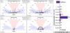
Outcome measuresThe primary outcomes measured were uncorrected distance visual acuity (UDVA), subjective refraction, corrected distance visual acuity (CDVA), and anterior segment evaluation using the SIRIUS (CSO) and anterior segment optical coherence tomography (MS-39, CSO).
The standard visual and refractive outcomes, as well as astigmatism outcomes have been analysed from postoperative day 1 to 12-month postoperative follow-up.
Statistical analysisData were analyzed using Excel (Microsoft Corp.). The significance of the differences was evaluated considering a metric distributed approximately as t with N-degrees of freedom, where N is the size of the sample as number of patients. Paired t-tests were used to determine statistically significant changes, with a P value of <0.05 considered significant.
Figures have been created using the mEYEstro and Astigmatic software packages.18,19
ResultsThe postoperative visual outcomes from day 1 to 1-year postoperative follow-ups after lenticule extraction in high myopia above −10D after a retrospective review chart including 49 eyes of 35 patients consecutively treated with the SCHWIND ATOS using lenticule extraction for high myopia above −10D who had at least 6-months follow-up completed are presented in Figure 1 using the standard visual and refractive outcomes, whereas Figure 2 presents astigmatism outcomes.

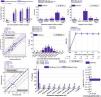
Standard graphs for reporting astigmatism outcomes of refractive surgery for 49 eyes consecutively treated for myopic defocus above −10D A) Cumulative Snellen visual acuity. B) Difference between achieved UDVA and pre-operative CDVA baseline. C) Change in Snellen lines of CDVA. D) Scattergram of the SEq. E) Predictability of the SEq respect to the target. F) Stability of the postoperative SEq. G) Predictability of the refractive astigmatism respect to the target. H) Scattergram of the astigmatic correction. I) Predictability of the refractive astigmatic axis respect to the target.

Uncorrected Distance Visual Acuity (UDVA): At the last follow-up, 73 % of the eyes achieved UDVA of 20/25 or better. Additionally, 88 % of the eyes had postoperative UDVA within 1 line of preoperative Corrected Distance Visual Acuity (CDVA).
Corrected Distance Visual Acuity (CDVA): Preoperatively, the mean CDVA was 20/22±6, ranging from 20/20 to 20/40. Postoperatively, over 94 % of the eyes reached same or gained lines of CDVA, with only 6 % of eyes losing 2 lines of CDVA.
Refractive outcomesSpherical Equivalent (SEq): The postoperative spherical equivalent was −0.07±0.55D, with 92 % of the cases within 0.75D of the target. The stability of SEq from 1-week to 1-year postoperative follow-ups was excellent, with only 10 % of eyes changing by 0.5D of defocus.
Refractive Astigmatism: Postoperative refractive astigmatism was 0.10±0.20D, with 96 % of the cases within 0.5D and 93 % within 5 degrees of the attempted axis. The astigmatic correction index was 1.02±0.28, with 76 % of the cases within 10 % deviation.
Predictability and stabilityPredictability: The scattergram for spherical equivalent showed a slight undercorrection, with 92 % of the eyes within 0.75D of the attempted SEq. The scattergram for astigmatic correction showed no overcorrection or undercorrection, with 96 % of the eyes having <0.5D of residual astigmatism and 93 % within 5 degrees of the attempted astigmatic axis.
Stability: The postoperative outcomes demonstrated excellent stability from 1-week to 1-year follow-ups, with minimal changes in defocus and astigmatism.
Intraoperative and postoperative complicationsIntraoperative: Lenticule removal was complete without relevant intraoperative complications in all patients.
Postoperative: At the last follow-up, 6 % of the eyes lost 2 lines of CDVA. However, 94 % of the eyes either maintained or gained lines of CDVA.
Corneal thickness and tissue alterationLenticule Thickness: The actual maximum lenticule thickness measured postoperatively ranged from 104 to 153 µm (130±15 µm).
Residual Stromal Thickness (RST): The actual RST ranged from 275 µm to 364 µm (317±31 µm), with total corneal thickness postoperatively from 392 µm to 513 µm (454±31 µm).
Percentage Tissue Altered (PTA): The PTA ranged from 36 % to 51 % (43 %±4 %).
DiscussionWe evaluated the postoperative visual outcomes from day 1 to 1-year postoperative follow-ups after lenticule extraction in high myopia above −10D. For this aim, a retrospective review chart identified 49 eyes of 35 patients treated with the SCHWIND ATOS using lenticule extraction for high myopia above −10D who had at least 6-months follow-up completed. For those, the standard visual and refractive outcomes, as well as astigmatism outcomes have been analysed. The most important paradigm for vector analysis was introduced by Alpins over 30 years ago and continues being the seminal work for analyzing astigmatic corrections.20
Retrospective studies may suffer from inadvertent selection bias (the problem of analyzing “selected” patients from the database), unless all patients qualifying to enter the study are included. This observational study involved the retrieval of consecutively treated eyes (meeting the high myopia definition used in the work). There were no other criteria than meeting the high myopia definition and having completed the required follow-up.
Despite the definition of high myopia to qualify for this work was −10D of myopia, two cases were treated for less than −10D. Actually, one patient was very close to −10D MRSE both eyes, since the author knows the system slightly overcorrects (∼0.5D), eyes were planned with a nomogram; which in this case led to an attempted SEq (Figure 1) of −9.5D in one eye and −9.88D in the other eye.
This investigation tries to evaluate the postoperative visual outcomes (in terms of stability and predictability) of a relatively new femtosecond platform (SCHWIND ATOS®) in patients with myopia above −10 diopters. Although the topic of the research is not new, it provides some interesting data in patients with particularly high myopia.
This study demonstrates that lenticule extraction using the SCHWIND ATOS system for high myopia above −10D is both safe and effective. The postoperative visual outcomes indicate that a significant majority of patients achieved excellent visual acuity, with 73 % of eyes reaching UDVA of 20/25 or better, with 59 % achieving 20/20 or better and 88 % within 1 line of preoperative CDVA, and only 6 % eyes losing 2 lines of CDVA. The refractive outcomes were also highly accurate (−0.07±0.55D for SEq and 0.10±0.20D for postoperative refractive astigmatism), with 92 % of cases achieving a postoperative spherical equivalent within 0.75D of the target and 96 % of cases achieving refractive astigmatism within 0.5D, with excellent stability from 1-week to 1-year postoperative follow-ups (with only 10 % eyes changing by 0.5D of defocus). Astigmatic correction index (1.02±0.28) showed 76 % of the cases within 10 % deviation.
The trends in LVC suggest a reduction of the range of refractive corrections treated using KLEx procedures, in favour of other intraocular procedures, such as phakic intraocular lenses (pIOL).3 This leads to a paucity of evidence in the high myopic subgroups, in particular beyond −10D of myopia.
For such high correction, not only visual outcomes and optical quality after KLEx but also continuous long-term monitoring for ectasia shall be performed, since long-term changes in visual quality and corneal stability require further investigation. Previous works on similar cohorts determined that KLEx is a safe way to correct for myopia higher than −10 D, with posterior corneal elevation (an early indicator of iatrogenic keratectasia) remaining stable 2 years after surgery. But also, a 4 years evaluation of the outcomes of KLEx for extreme high myopia and myopic astigmatism, suggested stable means of correcting high myopia and myopic astigmatism over a 4-year postoperative period.
The findings of this study are consistent with previous research on KLEx procedures for high myopia. Early studies have shown that KLEx can effectively correct myopia and myopic astigmatism over −10D, with stable posterior corneal surfaces and minimal long-term complications . This study further supports these findings by demonstrating excellent stability and predictability of refractive outcomes from 1-week to 1-year postoperative follow-ups.
SCHWIND ATOS is a relatively new development in femtosecond laser technology, and this is the first work specifically reporting on the high end of the correction ranges.
A feature of the SmartSight lenticule is the fact that it has a zero-thickness edge, and a lack of pedestal ensures that the removal is usually without risk of retained lenticule fragments. This suggests that the amount of tissue removal with SmartSight may be lower (both in thickness17 and volume21) compared to alternative KLEx procedures (including pedestal).
Cyclotorsion compensation is reported to provide an advantage with astigmatic values greater than 1.00 D.14 Since in this cohort 53 % of the treatments (26 eyes) had preoperatively treated refractive astigmatism above 1D, large cyclotorsion errors would have results in measurable residual astigmatism. The low impact of cyclotorsion, as demonstrated by 93 % of the eyes with angle of error withing 5° may be due to low torsional movements in this cohort along with proper cyclotorsion compensation from the system.
In this work only moderate OZs (5.5–6.2 mm, averaging 5.8 ± 0.2 mm) have been applied, which shall help keeping maximum lenticule thickness, residual stromal thickness, estimated total corneal thickness after extraction, and PTA within reasonable ranges. The results show that lenticule thickness was actually lower than planned (comparable to previous publications),22 whereas residual stromal thickness and estimated total corneal thickness after extraction were 20–30 µm thicker than expected (independently confirmed by other surgeons in personal communications, David Sung Yong Kang MD, unpublished data, written communication/email, September 12, 2024), both leading to smaller PTA than anticipated.
However, during the planning despite the use of moderately small OZs, all treatments were exceeding 40 % PTA during planning, whereas 70 % of the treatments actually exceeded 40 % PTA at the last follow-up. This reinforces the notion that continuous long-term monitoring for ectasia shall be performed. Further reducing cap thickness (currently limited to 100 µm minimum) may help reducing PTA while not negatively affecting visual outcomes.23
Counterintuitively, wider OZs (i.e., larger percentage volume altered, PVA)21 may not be more detrimental to the risk of iatrogenic ectasia (and might be actually protecting against it).24
In all patients, lenticule removal was complete without relevant intraoperative complications. Pre-operatively, the patients had a CDVA below the normal range (mean: 20/22±6; range: 20/20 to 20/40). This CDVA could be a characteristic specific to our patient population. In this subgroup, 6 % of the eyes (three eyes) lost Snellen lines of CDVA at the months post-operatively. At the last follow-up visit, 88 % of the eyes had UDVA within 1 line of preoperative CDVA baseline.
The scattergram of surgically induced astigmatism versus target-induced astigmatism showed no under-or overcorrection. Our findings seem to challenge the notion of and concerns about the undercorrection of the astigmatic component in lenticule extraction procedures.25 This challenge had been implied in previous reports,26 but cannot be confirmed in our results, in which residual astigmatism did not exceed 0.75 D, and 96 % of the eyes had a residual astigmatism up to 0.50 D
The change in manifest refractive astigmatism correlated well with the attempted values, suggesting that laser surgery could modify the corneal contour properly in moderately toric eyes. Therefore, it can be inferred that the planned goal of the treatment was met, even if patients were slightly undercorrected for refractive astigmatism. Patients may have been habituated to undercorrection of the cylinder in spectacles at the time of the pre-operative assessment, and thus accustomed to some residual astigmatism. This might have resulted in the treatment plans also being designed based on the “underrefracted” astigmatism. These results highlight the need for better ways to properly determine how much astigmatism should be incorporated into the treatment. It could be hypothesized, similar to hyperopic cases, not to overplan the treatment with the knowledge of a potential regression occurring post-operatively, but to instead push the refraction as much as possible. If so, then the cylinder correction chosen should be closest to the corneal toricity and compatible with best CDVA, analogous to the most positive SEQ compatible with best CDVA.27 In this regard, it also can be noted that patients may not accept high cylinder at the spectacle plane but may more easily do so on the corneal plane.
The outcomes reported here indirectly demonstrate proper placement of the correction profile in terms of both centration and cyclotorsion. We demonstrated that myopic astigmatism correction with SCHWIND ATOS SmartSight provided good results in terms of efficacy, safety, predictability, and visual outcomes in the first year of follow-up. In addition, the outcomes showed good predictability for both SEq and cylinder refraction. This finding is important and counters the accepted notion in the literature concerning undercorrections for sphere and cylinder in lenticule extraction, possibly because the treatment includes a transition zone and no lenticule side cut (thus no minimum lenticule thickness).
Currently, we are using low-energy asymmetric settings of 85 nJ in 5.5 × 2.8 µm distance (552 mJ/cm2 and ∼50 mW).
Laser refractive surgery aims to correct patients’ refractive error by removing a portion of corneal tissue respective to the aimed correction. Thus, it would be logical to think that deviations in the lenticule thickness (amount of tissue removal) would determine refractive deviations in the final outcomes. On the other hand, removing too much tissue may result in unstable corneas post-op (at least for those corneas at risk).
In the presence of strong epithelial remodeling,28 the reduction in stromal thickness may be a better predictor for the thickness of the removed lenticule. On the other hand, using stromal thickness may be prone to more noise since the segmentation of the epithelium-Bowman interface is clearly more difficult than that of the air-tear film (or tear film-epithelium).
Results show a lower reduction in CCT compared to laser read-out and the same has been reported for competing devices.29
Other groups assessed the accuracy and influence factors of predicting lenticule thickness in an alternative technique (SMILE using the VisuMax system by CZM, Jena, Germany).30 In general, lenticule thickness was also always lower than laser read-out.
However, opposite to our findings, refractive treatments were also undercorrected, correspondingly deviations in the lenticule thickness (amount of tissue removal) determine refractive deviations in the final outcomes. The extent of undersize in the thickness of the lenticules with the alternative technique was much higher than in this report, suggesting that may be the thickness deviations observed in this cohort may not be sufficiently large (despite being systematic) to be reflected in the residual post-operative refraction. Alternatively, the reduced lenticule thickness (while preserving the refractive correction) could be associated to a smaller than planned effective optical zone.
The planned OZ for SmartSight can be set freely between 5.5 and 8.0 mm diameter (+TZ of up to 0.8 mm outside of the OZ). Of course, as the lenticules get thicker (e.g., for higher corrections or larger optical zones), they are easier to extract, but less residual stroma is left behind. ATOS limits the maximum lenticule thickness between 25 and 165 µm. Further, ATOS imposes a minimum 275 µm calculated residual stromal thickness.
Corneas in Nepal are rather small, and so are pupil sizes as well; thus, a smaller OZ (compared to other regions in the world) is usually adopted. Further, treatments were in high myopia, which are associated with smaller planned OZ as a measure to reduce tissue consumption (and respect RST of 275 µm, with total corneal thickness after extraction above 350 µm).
The SCHWIND ATOS system's advanced femtosecond laser technology, including features such as zero-thickness edge lenticules and cyclotorsion compensation, contributed to the high accuracy and precision of the refractive corrections. The novel design of the SmartSight lenticule, which minimizes the risk of retained lenticule fragments, likely played a role in the smooth postoperative recovery and high visual acuity outcomes observed in this study.
So far, over 2200 patients completed the one-year follow-up, but not all had completed records or extreme myopia to be included in this chart review. All patients had a remaining corneal residual bed thickness of 275 µm or more. A complete dissection and removal of the lenticule was achieved in all cases without relevant intraoperative complications, and at post-operative day 1, all patients had a clear cornea.
The results of this study suggest that lenticule extraction using the SCHWIND ATOS system is a viable option for correcting high myopia above −10D. The high accuracy and stability of the refractive outcomes indicate that this procedure can provide predictable and reliable visual improvements for patients with extreme myopia. Additionally, the minimal intraoperative and postoperative complications observed in this study highlight the safety of the SCHWIND ATOS system.
This study has several limitations. Firstly, it is a retrospective review, which may introduce selection bias. Although the study included consecutively treated eyes, the inherent limitations of retrospective analyses should be considered. Secondly, the follow-up period was limited to 1 year, and longer-term outcomes beyond this period were not assessed. Future studies with extended follow-up periods are necessary to evaluate the long-term stability and safety of the procedure.
Further research is needed to explore the long-term outcomes of lenticule extraction using the SCHWIND ATOS system, particularly in terms of corneal stability and visual quality. Additionally, studies comparing the SCHWIND ATOS system with other femtosecond laser platforms for high myopia correction could provide valuable insights into the relative advantages and limitations of different technologies.
Lenticule extraction using the SCHWIND ATOS system for high myopia above −10D is a safe and effective procedure, demonstrating excellent visual and refractive outcomes with high predictability and stability. This study provides valuable data supporting the use of this novel femtosecond laser technology for extreme myopia correction, paving the way for future advancements in refractive surgery.
ConclusionsThis study demonstrates that lenticule extraction using the SCHWIND ATOS system for high myopia above −10D is both safe and effective. The procedure resulted in excellent visual and refractive outcomes, with high predictability and stability from 1-week to 1-year postoperative follow-ups. The majority of patients achieved significant improvements in uncorrected distance visual acuity (UDVA) and maintained or improved their corrected distance visual acuity (CDVA).
The SCHWIND ATOS system's advanced femtosecond laser technology, including features such as zero-thickness edge lenticules and cyclotorsion compensation, contributed to the high accuracy and precision of the refractive corrections. The novel design of the SmartSight lenticule minimized the risk of retained lenticule fragments, facilitating smooth postoperative recovery.
Despite the promising results, the study's retrospective nature and limited follow-up period highlight the need for further research. Future studies should focus on long-term outcomes and comparisons with other femtosecond laser platforms to provide a comprehensive understanding of the procedure's efficacy and safety.
We postulate that the advanced myopic ablation pattern of the low-energy asymmetric spacings results in a smoother extraction and thus a more rapid visual recovery for patients even for high myopic corrections.
In conclusion, lenticule extraction using the SCHWIND ATOS system represents a viable option for correcting high myopia above −10D, offering predictable and reliable visual improvements with minimal complications. This study provides valuable data supporting the use of this novel femtosecond laser technology for extreme myopia correction, paving the way for future advancements in refractive surgery.


