


This study explored whether retinoscopy (RET) provides comparable results of relative peripheral refraction (RPR) to open–field autorefractometry (AR) in myopic subjects.
MethodsPeripheral refraction was measured in 20 myopic and 20 control adult subjects. Both central and peripheral refraction (20° nasal and temporal eccentricity) were measured using RET and open-field AR. Differences in the median central spherical equivalent (SE), median RPR, and median J45/J180 power vectors between the RET and AR techniques were analyzed. Moreover, Bland – Altman plots were used to assess the agreement between RET and AR methods for RPR measurements in MG.
ResultsFor MG, the median RPR values were positive (hyperopic shift), and no significant differences were observed between the RET and AR techniques with respect to RPR measurement. In addition, we did not observe any significant differences in the RPR values between the nasal and temporal eccentricities for either the RET or AR technique for myopic subjects. There was also a significant correlation and agreement between the RET and AR technique for RPR measurements. With respect to central refraction, the median SE was slightly more positive for the RET than for the AR technique. Inside the CG, we also found significant correlation between the RET and AR technique for RPR measurements, and we observed a myopic shift in peripheral eccentricities.
ConclusionOur results show that retinoscopy may be a useful tool for objective measurements of RPR in myopic subjects and may be used interchangeably with the open-field AR method in everyday clinical practice.
It is generally accepted that peripheral defocus may play an important role in the process of eyeball elongation. Both animal and human studies have demonstrated that the sign of retinal defocus (hyperopic or myopic) can be recognized by the visual system and can influence the emmetropization process.1-4 Myopia is usually associated with a hyperopic relative peripheral refraction (RPR), whereas hyperopia or emmetropia is often associated with a non-myopic or myopic shift in the peripheral retina.4–9
Repeatable and accurate methods of both central and peripheral refraction examination are essential because most of the current approaches to myopia control (multifocal soft contact lenses, orthokeratology or defocus incorporated multiple segments (DIMS) spectacle lenses) are based on peripheral hyperopic defocus modification. Thus, knowledge of the peripheral refraction profile in myopic children and children at risk for developing myopia is essential in daily optometric or ophthalmic practice. Although retinoscopy has been already used to measure peripheral refraction, in practice, peripheral refraction measurements are usually performed for scientific research purposes using an relatively expensive open-field autorefraction technique. To the best of our knowledge, the first peripheral refraction measurements by retinoscopy (eccentric retinoscopy or off-axis retinoscopy) were conducted by Rempt et al. (1971)10 and the first link between peripheral defocus and myopia onset in humans was suggested by Hoogerheide et al. (1971).10,11 Since that time numerous human studies have shown that myopic eyes usually demonstrate a hyperopic defocus in the periphery.4,6–8,12 However, the results related to nasal – temporal asymmetry in RPR values showed rather conflicting results.
Radhakrishman et al., (2013)13 studied the peripheral refraction up to ±35° using an open-field AR and observed that all young myopic subjects (aged 14–22 years) showed a hyperopic RPR with the nasal retina exhibiting a slightly higher value of hyperopic RPR than the temporal retina, which may suggest that RPR levels may differ between the nasal and temporal eccentricities in myopic subjects. On the other hand, Yelagondula et al. (2022)14 measured the peripheral refraction using an open-field AR in 10° intervals up to ±30° and concluded that adult myopes showed an asymmetric type of peripheral refraction with relative hyperopic defocus in temporal retina and myopic defocus in the nasal retina. In addition, Atchison et al. (2006)7 did not observe any significant asymmetry in temporal-nasal peripheral refraction profile in young adult myopic subjects. Thus, further research are needed to verify whether adult myopes show symmetric or asymmetric peripheral refraction profile.
Unlike to open-field AR, retinoscopy is a relatively inexpensive method which allows to assess the state of refraction. Although several previous studies have shown that retinoscopy provides good agreement with the open-field autorefraction method with respect to central refraction or accommodative response results,15–18 to our current knowledge, there is a lack of studies that attempt to compare the agreement of the open-field autorefraction technique with retinoscopy with respect to RPR measurement in myopic subjects. From clinical point of view, it is important to know whether different methods (RET vs. AR) of RPR assessment in myopic subjects are interchangeable. Furthermore, an insight into the profile of peripheral defocus in children may be useful not only for predicting the risk of myopia onset but also for making a proper decision regarding the optimal method of myopia control (e.g. optical therapy for children with significant hyperopic defocus or pharmacological treatment for others). Therefore, it is important to accurately measure peripheral refraction and to know how different optical strategies dedicated to myopia control influence the peripheral refraction.
Thus, the aims of this study were to determine: (1) if retinoscopy results of RPR are comparable to open -field autorefraction technique, (2) if using retinoscopy makes it possible to detect temporal and nasal hyperopic RPR in myopic eyes, and (3) if nasal and temporal RPR show symmetric profile in myopic subjects
MethodsParticipantsForty adult subjects (mainly optometry students, eight males) took part in this study. The myopic group (MG) consisted of twenty subjects (mean age 28 ± 13years), and the control non-myopic group (CG) consisted of twenty subjects (mean age 24 ± 9 years). Myopia was defined as the spherical equivalent of ≤ - 0.50 diopters (D), emmetropia was defined as the spherical equivalent between −0.49 D and +1.00 D, whereas hyperopia was defined as the spherical equivalent of > +1.50 D, similarly as in other studies.9,19,20 MG consisted of subjects with myopia (mean spherical equivalent was −2.76 D, SD: 1.77, range from −0.50 to −6.75 D). In the CG eighteen subjects were emmetropes and two had low hyperopia, no more than +1.50 D (mean spherical equivalent was +0.36 D, SD: 0.45, range from −0.13 to +1.50 D). Based on a medical interview, all subjects were healthy, without any ophthalmic or general diseases, and none of them took drugs that might affect the visual system. Exclusion criteria were strabismus, amblyopia, nystagmus, reduced best-corrected visual acuity (unknown etiology) as well as astigmatism above 1.00 D. Each subject underwent optometric examination including visual acuity measurement using Snellen letter chart reading, presence and magnitude of ocular misalignment using prism cover test, basic binocular vision functions assessment with Worth four dot test, and subjective (if needed) and objective refraction measurement (central and peripheral) using retinoscopy and open-field autorefraction. All of the tests used, with the exception of open-field autorefractometry, are standard in optometric practices, and detailed information on the procedures are presented in the professional literature.21
In order to maintain a more natural visual condition and to avoid potential side effects of cycloplegic agents, all measurements were under non-cycloplegic conditions (neither mydriatic nor cycloplegic drugs were used) and each subject was allowed to blink during the measurements (as needed). It is worth to note, that peripheral retinoscopy is a challenging procedure due to increased aberrations at peripheral eccentricities. In clinical practice, the observed reflex becomes more distorted and the direction of retinal reflex movement becomes more difficult to define. Consequently, the neutralization endpoint becomes more difficult to precisely assess. Therefore, to reduce a potential error in the peripheral refraction results, we decided not to exceed 20° during the RPR examination. In addition, Moore et al., (2014)22 showed that the repeatability of peripheral open-field autorefraction measurements decreases with increasing eccentricity
Experimental procedureInitially, the central and then the peripheral horizontal refraction, at an angle of 20°, were measured by the use of retinoscopy (Heine Beta 200 Retinoscope) using Retinoscopy Rack Set, for both the right and left eye of each subject (86 eyes). Similarly as in previous studies, only right eyes were analyzed.7,9,23 During the retinoscopy procedure, the 0.05 (20/400) Snellen optotype was fixated at a distance of 4 m. The working distance was 50 cm and there were no fogging to control accommodation in the fellow eye, instead of this subjects were instructed to fixate a large (0.05 Snellen letter) nonaccommodative target at a distance of 4 m in a dimly lit conditions. It is worth to note, that several previous studies reported no significant difference between peripheral refraction measurements made with the eye turn and the head turn method. Subjects in this study were instructed to turn their eyes at the fixation target, with the head kept stationary. The fixation target was presented at either 20° temporal or 20° nasal in random order and an off-axis retinoscopy was performed. Similarly, central and peripheral refraction at an angle of 20° was measured with the open-field autorefractor (NVision – K 5001, Shin-Nippon). Since the consistent visual fixation is crucial for accurate peripheral refraction examination, subjects were instructed to carefully and stable fixate a target. In addition, the state of fixation was subjectively monitored by the examiner who also constantly reminded about stable fixation on the target. The measurements were taken only when the examiner was sure that the visual fixation was maintained on the target. Each measurement was taken three times and averaged. During the open-field autorefraction procedure, subjects were instructed to keep their heads stationary, with the chin and forehead resting firmly against the chin and forehead rest and to turn the eyes to fixate the peripheral target. The fixation target was a large red cross (18 × 18 cm) surrounded by a red border. In the center of the cross was a 2 × 2 cm white dot, which provided a fixation target. The stimulus for fixation was supplied by the AR manufacturer and the pattern used provided a stable stimulus for fixation and accommodation.
The research was approved by the Ethics Committee of Adam Mickiewicz University in Poznan, Poland (No.: 27/2017/2018) and was conducted in accordance with the tenets of the Declaration of Helsinki. Informed consent was obtained from all participants.
Statistical analysis and definitionsStatistical analyses were performed with Statistica 10 software (StatSoft) and MedCalc software. Only results from the right eyes were analyzed. Since not all data had normal distribution, non-parametric tests were used. Differences in the median spherical equivalent (SE), median RPR, and J45/J180 indexes between methods were tested with the Wilcoxon test. The U Mann-Whitney test was used to compare median RPR between groups and Friedmann Anova was used to evaluate J180 and J45 profiles. Spearman correlation was used to evaluate relationships between AR and RET methods. The level of significance was P <= 0.05. Since, the differences between the nasal and temporal RPR values were normally or nearly normally distributed, Bland – Altman analysis was used to assess the agreement between two measurements methods: RET and AR.24
For statistical analysis, any spherocylindrical refractive error obtained from the RET method and AR was converted into power vectors according to the formula:
where SE is the spherical equivalent, S is the spherical power, C is the negative cylindrical power, α is the cylindrical axis, and J180 and J45 are the power of the two Jackson cross-cylinder components. A positive value of J180 indicates with-the-rule (WTR) astigmatism, whereas a negative value of J180 indicates against-the-rule (ATR) astigmatism. J45 refers to a cross-cylinder set at 45° and 135°, representing oblique astigmatism. The negative value of J45 indicates that the negative cylinder axis lies between 90° and 180°, whereas a positive value indicates that the negative cylinder axis lies between 0° and 90°. Power vectors are geometrical representations of spherocylindrical refraction in 3 basic dioptric components, which are mathematically independent of the others.25


The absolute peripheral refraction at a given eccentricity (20° nasal or 20° temporal) was the spherical equivalent (SE) at that eccentricity. The relative peripheral refraction (RPR) at a given eccentricity was calculated according to the formula SE at that eccentricity minus central SE. Thus, a positive value of this formula indicates that relative peripheral hyperopia (RPH) exists, whereas a negative value indicates relative peripheral myopia (RPM).
ResultsAs can be seen in Table 1, in CG median SE values in the central retina measured with AR and RET methods were equal (+0.25, P=.72) and in MG there was a slight shift in a more myopic direction for RET than AR (−2.57 vs. −2.56, P=.05, for RET and AR, respectively). There was also a very strong positive correlation in the value of SE between both methods for all analyzed eyes (R = 0.98, P<.001) as well as for MG (R = 0.99, P<.001) and for CG (R = 0.87, P<.001).
Median values of spherical equivalent of refractive error (SE) measured in the central retina (CR) for myopic (MG) and control (CG) groups. AR – autorefractometry method, RET – retinoscopy; O1 and Q3 – lower and higher quartile.
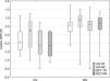
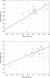
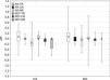
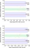
The values of RPR are presented in Table 2 and in Figure 1. As can be seen, RPR in MG was positive (hyperopic defocus) in temporal and nasal eccentricity, both for AR and RET methods (Figure 1). When the temporal area was analyzed, the median value of RPR measured with the RET method was slightly more positive than with AR (+0.62 D vs. +0.56 D, for RET and AR respectively) but this difference was statistically insignificant (P=.586). In the nasal direction, a slightly more positive value of RPR with the AR (Figure 1 left, MG) method was obtained when compared to the RET (Figure 1 right, MG) method (+0.87 vs. +0.69 D, for AR and RET, respectively) but again, this difference was statistically insignificant (p=.522). There was also a significant positive correlation between RET and AR methods (Figure 2) in RPR for MG measured in the temporal retinal (Figure 2, upper) direction (R = 0.60, P=.005) but less positive correlation was observed in the nasal direction of the retina (Figure 2, lower) (R = 0.42, P=.064).
Median values of relative peripheral refraction (RPR) for temporal (TR), and nasal (NR) retina, for myopic (MG), and control (CG) groups. AR – autorefractometry method, RET – retinoscopy; O1 and Q3 – lower and higher quartile.


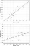
More differences were found in CG. Here, again significant correlations between methods in RPR measured in the temporal (Figure 3) direction (R = 0.86, P<.001) as well as in the nasal (Figure 3) retinal area (R = 0.56, P=.010) were found. However, when median RPRs were compared between both methods used, little shift in the negative direction with the RET method compared to AR was found (RPRTR: −0.62 vs. −0.44 D, for RET and AR, respectively, P=.018; RPRNR: −0.62 vs. +0,25 D, for RET and AR, respectively, P=.002) (Figure 1, CG).

Inside the MG, we did not observe any significant differences in RPR between the nasal and temporal retina, both for the RET (+0.62 D RPRTR vs. +0.69 D RPRNR, P=.588) as well as the AR method (+0.56 D RPRTR vs. +0.87 D RPRNR, P=.420). Inside the CG, statistical analysis showed significant differences between the RPR in the temporal and nasal retina only for the AR method (−0.44 D RPRTR vs. +0.25 D RPRNR, P=.025) but not for the RET method (−0.62 D RPRTR vs. −0.62 D RPRNR, P=.627).
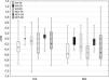
J180 and J45 vector valuesFor the vector analysis, we converted the spherocylindrical values to power vector notations. J180 vector values are demonstrated in Table 3 and Figure 4. The value of this power vector was dependent on the eccentricity. For MG, there was a statistically significant negative increase in J180 with eccentricity, for both the nasal and temporal retina, indicating a trend toward against-the-rule astigmatism in the horizontal meridian with increasing eccentricity. This observation was true with the AR (Figure 4 top left corner, MG) method (χ2(20,2)=14.40, P<.001) as well as with the RET (Figure 4 top right corner, MG) method (χ2(20,2)=10.22, P=.006). Similar findings occurred for CG using the AR (Figure 4 top left corner, CG) method (χ2(20,2)=11.10, P=.004) but with the RET (Figure 4 top right corner, CG) method, differences between the central and peripheral J180 were only close to significant (χ2(20,2)=4.91, P=.086). No difference in the values of J180 was found between both methods used in any eccentricity for MG (Table 3, P>.499), or for CG (Table 3, P>.351).
Median values of J45 and J180 indexes for temporal (TR), central (CR), and nasal (NR) retina, for myopic (MG) and control (CG) groups. AR – autorefractometry method, RET – retinoscopy; O1 and Q3 – lower and higher quartile.

J45 vector values are presented in Table 3 and Figure 5. No significant difference in J45 for MG between different retinal eccentricities was noted when AR was used (χ2(20,2)=1.33, P=.513) or with the RET method (χ2(20,2)=0.48, P=.786). Similarly, the profile of J45 was flat for CG when refraction was measured with AR (χ2(20,2)=5.85, P=.054) as well as with the RET method (χ2(20,2)=4.75, P=.093). Again, no significant difference in the J45 index was found between both methods used when MG (Table 3, P>.365) and CG (Table 3, P>.063) were analyzed. Mean differences between the RET and AR methods with respect to RPR in MG are presented in Bland-Altman plots (Figure 6). The Bland-Altman method of comparison showed that the 95 % Limits of Agreement (LoA) between the RET and AR for nasal and temporal RPR was 0.94 to −1.19 and 1.12 to −1.33 respectively (Figure 6).


Bland-Altman plots showing good agreement between RET and AR for Nasal (upper) and Temporal (lower) RPR measurements in MG. The solid blue line represents the mean difference. The limits of agreement (LoA) are defined as the mean difference ± 1.96 SD of differences (upper and lower limits are shown as red dotted lines), 95 % Confidence Intervals (CI) for mean difference and agreement limits are shown as green and blue shaded areas respectively.
The primary aim of this study was to determine whether retinoscopy can be a useful and reliable method of relative peripheral refraction (RPR) measurement in myopic subjects. To be specific, this study explores three questions: (1) Does retinoscopy provide comparable results for central refraction as well as RPR with the open-field autorefraction method? (2) Do adult myopic subjects show a hyperopic shift in the peripheral (20° nasal and temporal) retina ? (3) Does RPR show comparable values in the nasal and temporal retina or is there a significant asymmetry between the nasal and temporal eccentricities in myopic subjects?
Regarding the central refraction measurements, we did not observe any significant differences between the retinoscopy and open-field autorefraction under non-cycloplegic conditions. This was not surprising, and our results are in good agreement with previous studies.15
With respect to RPR in the myopic group, we observed a typical small hyperopic shift in both the nasal and temporal peripheral retina (both for RET and AR methods). Our findings suggest that both retinoscopy and open-field AR may be useful tools for objective measurements of RPR, providing comparable results and good agreement. Positive median values of RPR found in MG was more than +0.50 D and less than +1.0 D, what is in a good agreement with previous studies on peripheral defocus in myopic subjects (usually not exceeding +0,75 D).4,7,22,26 In addition, we did not observe any significant difference between the nasal and temporal eccentricity with respect to median RPR, for both RET and AR methods. Zheng et al. (2022)26 studied the associations between regional peripheral refraction and myopia development in young subjects aged 18–28 years and concluded that eccentricities between 20°- 35° may be closely related to central refraction development and eye growth regulation. Since hyperopic stimulation of peripheral retina may play and important role in the onset and development of myopia, it seems that 20° off-axis retinoscopy may provide valuable information about RPR and may facilitate a decision-making process related to optimal method of myopia progression control.
When RPR for the control group was analyzed, a small myopic defocus in the nasal and temporal retinal eccentricities was found (especially for the RET method). To our surprise, a small hyperopic RPR (median: +0.25 D) in CG was observed only in the nasal retina when AR method was used but these values are within the measurement error range of the AR device. Furthermore, we cannot exclude the effect of decreased repeatability of peripheral autorefraction measurements with increasing eccentricity.22 In general, the results related to RPR in emmetropic or hyperopic eyes are in a good agreement with other studies.4,6–8
Our findings related to power vector analysis showed a significant negative increase of J180 in MG for peripheral areas, suggesting a trend toward against-the-rule astigmatism in myopic subjects both for nasal and temporal horizontal eccentricities. This was true for both RET and AR methods, with no significant differences between either method. Similar findings related to J180 power vector (a shift toward against the rule astigmatism with increasing eccentricity) were observed for CG using the AR method (with the RET method, differences between the central and peripheral J180 values were close to significant in CG). Our findings are in a good agreement with Chen et al. (2010) study, where also increasing negative values of J180 power vector in the horizontal meridian with increasing eccentricity was found in both myopic, emmetropic and low hypermetropic subjects.23
With respect to the J45 component in MG and CG, we did not observe any significant differences between the central, nasal, or temporal retinal eccentricities, which suggests there is no significant trend toward oblique astigmatism in the horizontal peripheral (20°) retina in our myopic as well as control subjects.
Peripheral refraction measurements seems to be an important part of in-office examination since almost all optical methods are based on the assumption that peripheral hyperopic defocus should be reduced, and even myopic shift in refraction should be induced in subjects with progressive myopia. This can be done with orthokeratology,27 multifocal or bifocal contact lenses,28–30 or with novel design spectacle lenses.31 In addition, it was shown that baseline RPR may affect myopia control effect in children wearing Defocus Incorporated Multiple Segments (DIMS) spectacle lenses.32
Thus, accurate, quick, reliable and inexpensive method of peripheral refraction examination looks to be important when optical methods for controlling the progression of myopia are considered. Since multifocal soft contact lenses with high addition dedicated to myopia control may affect some visual functions,19,20,33 it seems to be reasonable to examine the RPR value before their application in order to adjust the optimal peripheral defocus in subjects with progressive myopia. Thus, when the application of any of the myopic control methods is considered, it is recommended to measure RPR and to check whether the applied intervention really changed the peripheral defocus. Since not all myopic subjects manifest peripheral hyperopic defocus, the ability to measure RPR in everyday practice may provide clinically relevant information in the context of customizing optical interventions for patients with progressive myopia. Methods such as orthokeratology, multifocal contact lenses or DIMS-type spectacles reduce the hyperopic defocus on the peripheral retina. They are therefore suitable for patients with hyperopic defocus, but not all types of myopia show this characteristic of the retinal image in the eye. Retinoscopy allows for a quick assessment of the degree of peripheral defocus and make an informed decision as to whether these optical methods are indicated for the patient or whether pharmacologic therapy should be considered.
RET seems to be an attractive and available option to testing RPR as compared with open-filed autorefractometry. Although RET requires some experience, our results indicate that it can be a valuable method of RPR examination and can be implemented not only in adults but also in myopic children. We are aware that this study has several limitations: the studied group was relatively small, there was no investigation of the effect of different degrees of myopia on RPR values, the peripheral refraction measurements were performed only at the angle of 20° by the same examiner, there wasn't investigation of repeatability, the studied groups included only adult subjects and there were no comparison between the cycloplegic and non-cycloplegic conditions. In addition, two subjects in our control group had low hyperopia. However, they manifested typical myopic shift with respect to RPR measurements and we decided not to exclude them from further analysis. Future research is needed to investigate the utility of retinoscopy to assess RPR in myopic children, to explore the effect of different degrees of myopia and astigmatism on RPR or to investigate the repeatability of RPR measurements across larger eccentricities using retinoscopy. Several previous studies showed that patients with high and moderate myopia had relative hyperopia at all eccentricities, whereas patients with low myopia and emmetropia had relative hyperopia only beyond 30 and 35° eccentricity, respectively. Thus, future studies are needed to assess the utility of retinoscopy to examine the peripheral refraction beyond the 20°.
ConclusionsThis study demonstrated that retinoscopy provides comparable results of relative peripheral refraction to open-field autorefraction in myopic eyes. To the best of our knowledge, this study is the first to compare RET and open-field AR methods with respect to RPR measurements. Although retinoscopy requires patience and is a challenging task, our findings provide further evidence that it can be used interchangeably with the open – field AR technique when there is a need to examine peripheral refraction or RPR in myopic subjects. This interchangeability of retinoscopy and open-field AR enhances the practical options available for measuring RPR in myopic subjects in everyday optometric or ophthalmic practice. We hope that our findings may help practitioners choose the most appropriate and available method for measuring RPR in their specific clinical practice. It is worth to note that retinoscopy has a several advantages compared to open-field AR such as cost-effectiveness, wider availability, mobility or more natural free viewing conditions. Our findings may also have potential broader implications where peripheral refraction examination is important and beneficial for patients, such as low vison subjects, neuro-optometric rehabilitation or retinal diseases such as age related macular degeneration. Considering the limitations of our study, future research is needed to explore the interchangeability of retinoscopy and open-field AR in different age groups, different degrees of myopia, different retinal eccentricities or in more complex clinical scenarios where peripheral refraction assessment is also important. It seems that after a short training under the supervision of an experienced specialist, peripheral retinoscopy could be implemented in clinical practice. Although in this study peripheral refraction was measured with the eye turn method, in practice one can also quickly measure the peripheral refraction by rotating the refraction equipment (retinoscope) around the fixed eye and head of the patients.


