
Evaluations of tear functions frequently involve some form of voluntary control over blink behaviour. To the degree that voluntary control of blinking risks departure from normal-range spontaneous blinking, the tear function findings from such studies may be confounded. Even subject awareness that blinking is being assessed may influence findings if such awareness results in any degree of voluntary control. Ideally, the influence on blink rate and tear functions induced by therapeutic or experimental interventions could be measured against a normal-range baseline spontaneous blink rate in order that any differences found could be validly attributed to those interventions. Sometimes pre-intervention ‘rest-related’ baseline blink rates have been incorrectly described as ‘basal’ blink rates without specification of pre-intervention conditions of ‘rest’ or consideration of any contributions from voluntary control. Also, studies which use only blink rates to measure blink efficiency ignore the critically important contribution of incomplete blinking to blink inefficiency. This review finds that the assessment of normal-range spontaneous blink rates depends on measurement conditions which have frequently been ignored previously. For example, normal-range spontaneous blink rates appear more likely to occur with fixation targets which have a disengaged affect and an associated neutral influence on and from dopamine activity. Ideally, fixation targets should also involve minimal cognitive loading and vision demands. In addition, normal-range (symptom free) spontaneous blink rates are more likely to be assessed in a comfortable ambient environment without subject awareness that blink behaviour is being assessed and when voluntary blinking is not involved.
Las valoraciones de las funciones de la lágrima incluyen alguna forma de control voluntario del parpadeo. Hasta el punto de que el control voluntario del parpadeo puede tener su origen en el parpadeo espontáneo de rango normal, los hallazgos sobre función de la lágrima de dichos estudios pueden resultar confusos. Incluso la concienciación del sujeto acerca de que se está evaluando el parpadeo puede influir en los hallazgos, cuando dicha concienciación deriva en cualquier grado de control voluntario. De forma ideal, la influencia sobre las tasas de parpadeo y las funciones de la lágrima inducidas por intervenciones terapéuticas o experimentales podría medirse frente a una tasa de parpadeo espontáneo basal de rango normal, a fin de poder atribuir válidamente cualesquiera diferencias a dichas intervenciones. A veces, las tasas de parpadeo de referencia "relacionadas con el descanso" pre-intervención se han descrito incorrectamente como tasas de parpadeo "basal", sin especificar las condiciones "de descanso" pre-intervención, o la consideración de cualquier contribución del control voluntario. De igual modo, los estudios que utilizan únicamente tasas de parpadeo para medir la eficiencia del parpadeo, ignoran la contribución críticamente importante del parpadeo incompleto sobre la ineficiencia del mismo. Esta revisión encuentra que la valoración de las tasas de parpadeo espontáneo de rango normal depende de las condiciones de medición, que con frecuencia han sido ignoradas previamente. Por ejemplo, es más probable que se produzcan tasas de parpadeo espontáneo de rango normal con objetivos de fijación, que tienen un efecto de desactivación y una influencia neutra asociada sobre la actividad de la dopamina. De manera ideal, los objetivos de fijación deberían implicar también una carga cognitiva mínima y unas demandas de visión. Además, es más probable que las tasas de parpadeo espontáneo de rango normal (con ausencia de síntomas) se valoren en un entorno confortable, sin que el sujeto sea consciente de que se está valorando el parpadeo, y cuando el parpadeo voluntario no se ve implicado.
Blinking plays an important role in the maintenance of the integrity of the ocular surface because it contributes to the maintenance of ocular surface humidity and favours tear drainage, with expression and dispersion of lipids from meibomian glands.1 Spontaneous blinks are usually the most common form of blinking and have a major role in maintaining the integrity of the ocular surface by spreading and restoring the tear film thickness2 as well as maintaining tear layer stability as the basis for clear vision.3 Spontaneous blinking is an unconscious brief eyelid closure of both upper eyelids which occurs in the absence of any evident stimulus.3 Given that the mechanisms for the control of spontaneous blinking are not completely understood,3 the conditions of ‘rest’ under which spontaneous blink rates (SBRs) approach normal-range physiological levels cannot be accurately specified. Usually ‘basal’ is a descriptive for the lowest physiological level but as discussed further below, in the case of blink rates (BRs), factors such as dopamine activity and complex visual and/or cognitive demand can reduce BR to below normal-range SBR levels that might be associated with ‘rest’. As conditions for a state of ‘rest’ vary so might the associated SBRs be expected to vary. Reflex and voluntarily controlled blinking are the antithesis of spontaneous blinking but being able to confidently eliminate reflex and voluntary influences on BRs may sometimes not be possible. The rate and degree of tear film evaporation and cooling, or extent of tear film break up (TFBU) and the associated threshold level for increased osmolarity and dry eye symptom influence on BR, do not appear to be known. For example, threshold levels for symptoms due to hyperosmolarity to stimulate reflex blinking may vary with ocular surface sensitivity and the area of any TFBU for example.
Spontaneous or reflex or voluntary blinksSymptoms of dry eye disease (DED) are known to increase BRs.,4,5 Apparently, at some stage of DED progression, stimuli related to evaporative cooling and/or increased hyperosmolarity increase toward a conscious level and blinking becomes reflex. The demand for blink-related ocular surface maintenance becomes more critical according to the development of one or more age-and/or pathology-related tear dysfunctions as well as in response to adverse ambient conditions and influences from environmental factors such as increased cognitive and visual demands. Under most normal-range circumstances and conditions, BRs not only vary according to a wide variety of such factors but also in response to the methods used to measure them. Examples of clinical and experimental methods for blink and/or tear function assessments which have involved various forms of voluntary control of blinking are shown in Table 1.6–11 For example, just before tear meniscus measurements, Qiu and co-authors asked each participant “to hold their blink during the acquisition of the optical coherence tomography scan and then they were instructed to blink normally”.6 An instruction to blink normally may result in voluntary blinking which alters tear meniscus volume for example. The characteristics of the blink prior to a scan, and any effect it has on tear meniscus volume is uncertain and, as a result, repeated scans involving voluntary blinks may not represent normal-range values.
Examples of instructions provided during clinical and experimental studies of tear functions which appear likely to induce voluntary blinking or other form of departure from normal-range spontaneous blink behaviour.
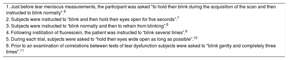
| 1. Just before tear meniscus measurements, the participant was asked "to hold their blink during the acquisition of the scan and then instructed to blink normally".6 |
| 2. Subjects were instructed to “blink and then hold their eyes open for five seconds”.7 |
| 3. Subjects were instructed to “blink normally and then to refrain from blinking”.8 |
| 4. Following instillation of fluorescein, the patient was instructed to “blink several times”.9 |
| 5. During each trial, subjects were asked to “hold their eyes wide open as long as possible”.10 |
| 6. Prior to an examination of correlations between tests of tear dysfunction subjects were asked to "blink gently and completely three times".11 |
Compared to conditions for SBRs, other characteristics of blinks may vary widely under voluntary control. Accordingly, voluntary control could be invasive with respect to normal-range spontaneous blink performance; perhaps especially when effort is made to be sure that a full blink is achieved. For example, voluntary blinks may be more forceful than spontaneous blinks and so more likely to promote lipid secretion and increase tear layer thickness and tear meniscus volume. Voluntary blinks involving greater force could also promote faster tear drainage or create tear surface irregularities.
The evidence that BR increases as a function of time on task is compelling but apart from the effects of fatigue, for example, other environmental variables influencing BR include the complexity and associated acuity demands of concurrent visual activity as well as cognitive factors.12 For example, cognitive factors include for instance, the perceptual demand of the concurrent activity such as blink timing (interblink intervals) with respect to information processing requirements.12 Acosta and co-authors showed how activities such as listening and talking can increase BRs while tasks involving visual information processing such as reading, can reduce BRs.1 Some other influences on BR have been broadly described as attention, level of concentration, as well as stress and anxiety.13 A videotape recording of BR while subjects were either resting quietly, reading aloud or conversing, led to the conclusion that cognitive processes were the chief influence on BR.14 Some of the variable conditions for ‘resting quietly’ in relation to an associated BR are elaborated below. BR inhibition during visual tasks has been assumed to preserve input information processing, thus reflecting attentional engagement.15 For example, visual activities involving a sequence of increasing degrees of attentional engagement such as looking straight ahead, watching a movie, identifying rapidly changing letters and playing a computer game, were found to progressively reduce BRs and elevate incomplete blinking rates in both dry DED and normal subjects.16
Blink efficiencyBlink efficiency is a measure of the ocular surface protective functions of blinking and is determined by the degree that blinking supports tear functions and so contributes to the health of the ocular surface. For example, both incomplete blinking and low rates of complete blinking reduce blink efficiency. Spontaneous blink efficiency is commonly assessed by only the measurement of BR,13,14,17,18 but several other features of blink behaviour appear likely to also contribute to blink efficiency. Variations in interblink intervals and different forms of incomplete blinking such as variations in blink amplitude (or degree of closure), the frequency of partial closures such as twitch blinks involving a very minor (aborted) degree of closure, flurries which are a series of rapidly repeated twitches, as well as differences in the speed of closure and opening phases for example,19,20 Variation in interblink intervals may be an indication of changing cognitive processing demands during assessment of BRs.12 Tear osmolarity increases with tear evaporation during an interblink interval. BR and completeness must be maintained at rates which prevents the formation of areas of significant evaporation and tear hyperosmolarity which have the potential to cause symptoms, tissue damage and/or a reduction in vision20 with associated reflex blinking.
Current knowledge of blink controlKnowledge about the control of spontaneous blinking is incomplete3 with little known about its neural basis.21 A simple part-explanation for complex blink behaviour is that the spinal trigeminal complex is a critical element in the generation of spontaneous blinks.21 However, BRs are otherwise believed to reflect a complex interaction between peripheral influences mediated by the ocular surface and the level of central dopamine activity,3 both of which are discussed further below. In order that an evaluation of tear functions is able to produce evidence that is representative of a patient’s customary blink behaviour, such evaluations should ideally involve a blinking phase which is representative of normal-range spontaneous blink activity. In this way the findings are more likely to have relevance to typical levels of blink efficiency for example. Rather than representing ‘background noise’ or being a randomly occurring phenomenon, variations in blood pressure have been shown to be the result of complex interactions between extrinsic environmental and behavioural factors and intrinsic cardiovascular regulatory mechanisms.22 To limit short-term influences on findings, suggested recommendations for the assessment of baseline blood pressure include rest in a comfortable physical position in a calm mental state for at least 5 min and no smoking or alcohol/caffeine intake for at least 30 min prior to assessment for example.22 Such measurements could provide a level of blood pressure against which the influence of an intervention could be compared. Similarly, the influence of an experimental intervention on blink activity would ideally be measured against normal-range baseline spontaneous blink performance. For example, assessment of tear film break up time (TFBUT) following voluntary complete blinking appears likely to thicken tear layers and give findings which are not representative of TFBUT during normal-range spontaneous blink activity.23 This review examines how those kinds of evaluations might and might not be validly achieved. PubMed searches (17th March 2019) using the terms spontaneous blink, voluntary blink, and reflex blink yielded 424, 190, and 4667 potentially useful publications respectively. Selections of those which were found to be the most relevant and representative of a balanced account of this topic, as well as some selected reports referenced in those publications, were included in this review.
Some differences between spontaneous, voluntary and reflex blinkingSpontaneous blinks are entirely sub-cortical while voluntary blinks are preceded by pre-motor cortical readiness.20 Voluntary blinks can differ from spontaneous blinks in regard to factors such as rate or interblink interval variability, degree of completeness, duration of closure, and the force involved, because they are associated with motor function.20 Consequently, the influences on tear functions can be very different according to the type of blink. The neural pathways involved in ocular surface controls over reflex blinking arise in ocular surface sensory input through the trigeminal nerve that projects to the motor neuron of the seventh cranial nerve.5 In this way, stimulation of the ocular surface, such as the symptoms associated with DED, increase both BR and regularity.5 Thus, when driven by symptoms, blinking in DED appears to be reflex rather than spontaneous. When task concentration levels were ‘controlled’, pneumatic stimulation of the ocular surface was found to increase BR and regularity in healthy human subjects.5 Apparently, the pneumatic stimulation and associated faster evaporation induced an artificial state similar to one that could be found in DED. The relationship between pneumatic stimulation and BR was linear suggesting a linear dose-related response relationship between ocular surface input and blinking.5 Even a barely noticeable air stimulus to the ocular surface, using a small electric fan at an average distance of 50 cm, produced an increased frequency and regularity of blinking.24 Apart from air movement, any other aspect of ambient conditions such as temperature and humidity could influence BR by altering the rate of tear evaporation. The input for triggering ocular surface-stimulated blinks depends on the integrity of the surface nerves.5 Naase and co-authors found that in a sample of 40 males without significant ocular surface disease, 37/40 (92.5%) showed a reduction in BR with topical ocular anaesthesia. For the 37 who showed a reduction, the mean reduction in BR was 37.4%.25 Loss of corneal sensitivity with age could be expected to influence BR.13 However, Bentivoglio and co-authors did not find any correlation between BR and age,14 and Sun and co-authors found a modest increase in BR with age.26 However, apart from corneal sensitivity, many other factors may confound the relationship between age, and BRs. It remains possible that increased BRs due to age-related DED could sometimes be moderated by concurrent reduced corneal sensitivity. The possibility that any sensory message sent by ocular surface cold receptors during evaporation also contributes to the modulation of blinking, is a functional alternative that deserves experimental scrutiny.27 Is it possible that normal-range evaporative increases in osmolarity during an interblink interval, could also provide stimuli for spontaneous blinking in healthy eyes? Such peripheral controls could occur in the absence of any apparent stimulus and could be independent of any central control mechanism. However, increased BR in DED4,5 may be due to faster evaporation and an associated more rapid rate of tear cooling and osmolarity elevation with resulting greater stimulation of the ocular surface. Even in healthy young females, a significant correlation between TFBUT and BR was found.13
Other influences on blink efficiencyDrew reported that BR varied inversely with task difficulty for all subjects that were studied.28 Variability of interblink intervals in normal subjects suggests an ability to vary BR and even suppress blinking according to variable perceptual/cognitive needs over time.20 Usually, TFBU does not occur during interblink intervals unless one or more factors such as incomplete blinking,29 tear layer instability, adverse ambient conditions, high visual acuity demand and/or elevated cognitive task loading are present. A dry eye subject’s need for more frequent blinking supersedes the requirements of a reading task for example.20 Again, such blinking is not spontaneous to the extent that factors influencing BR, such as comfort levels, are outside normal ranges. For individual subjects, BRs are specific to the circumstances under which they are measured and their individual responses to them. For example, the BR for 67.3% of healthy volunteers was greatest during conversation compared with while ‘resting quietly’, but then it was slower again when reading.14 However, for 22.7% of the same sample of healthy volunteers, compared to conversing the rates were fastest for resting although still slowest for reading.14 These findings suggest significant variation in individual normal responses. The level of cognitive activity during all three tasks for example may vary widely between individuals, and explain some of the differences in response, even during a so-called ‘resting’ period. The conditions for resting were not specified in that study.14 For example, thoughts may be dominated by rest disturbing stresses and anxieties, or some subjects may be familiar with yogic or other relaxation techniques and take the opportunity to achieve a deep level of relaxation during rest. The level of rest achieved may also vary with, for example, uncontrolled influences from visual activity and levels of body comfort.
Basal or baseline blink rate?BRs measured by observations from a video-recording after a 10-minute period of adaptation to unspecified experimental conditions of ‘rest’ and prior to exposure to experimental interventions, were described as basal BRs.30 Basal BR suggests a rest-related physiological SBR but, given that there are many factors that might be influencing BR during a period of ‘rest’, blinking prior to intervention might be more appropriately described as baseline rather than basal. Conditions such as subjects being completely rested before and during assessment and freedom from emotional stress, such as measurement-related apprehension,31 may be important. For example, ideally subjects would be unaware that they were being filmed or videotaped during a study of blink behaviour32 so that any related apprehension, anticipatory thoughts or other potential influences on BR, such as voluntary control of blinking, are avoided. Bentivoglio and co-authors described BRs recorded by videotaping during quiet ‘rest’ as the basal BRs when compared to those recorded during the experimental conditions involving reading and conversation.14 In that study, subjects were sitting and had agreed to being videotaped but were not aware of the purpose of the study.14 No other conditions of quiet rest were specified for that study and again, rather than basal, baseline might have been a more accurate description of the pre-intervention BR. To the extent that spontaneous blinks occur in the absence of any apparent stimulus,3 specification of measurement conditions would better inform baseline BR findings and their relationship to spontaneous-range BRs. Lack of awareness that blink behaviour is being examined might be a key feature of patient/subject ability to avoid voluntary blinking control. It is unknown to what extent would being physically uncomfortable and/or being exposed to unfamiliar measurement methods or equipment help to determine BR. For example, estimates of BR by observation during a biomicroscopic examination, which includes a potentially uncomfortable unnatural posture and mental state as well as abnormal lighting conditions, appear unlikely to be relevant to most real-world conditions.
Consequences of other aspects of methods of measurementCruz and co-authors reviewed BR assessment methods which included procedures such as lever arms being attached to the lid to detect lid motion, electrooculography, electromyography, wearing lid movement detecting spectacles as well as evaluation of high-speed videotapes.3 A magnetic search coil method was described as the gold standard method.3 However, that method requires that a small coil be taped to the upper lid and for the subject to be placed inside a magnetic field produced by a surrounding cubic frame.3 The high potential for invasiveness of these kinds of circumstances may reduce the possibility of capturing blink performance that is relevant to normal range real-world conditions.
Dopamine influences on blinkingDopamine is a hormone functioning as a neurotransmitter in several distinct systems within the brain.17 One such system plays a major role in reward-motivated behaviour with most types of reward increasing the level of dopamine in the brain.17 BR is affected by central dopamine level5 such that, in healthy adults, SBR is regarded as a marker of the level of central dopamine functioning33 and the daily pattern of SBR is a peripheral measure of central dopamine activity.18 The validity of these relationships is conditional on the degree that measured BR is influenced by factors other than dopamine. Blinks are only an indirect measure of dopamine level which appears to have an important role in cognitive control34 which, in turn, has a reciprocal influence on BR.12 The idea of an effective modulation of cognitive control through altered dopamine activity converges with common theories that suggest links between positive affect, dopamine and cognitive control.35 It has been assumed that the cognitive effects of positive affect are modulated by increased brain dopamine levels in the prefrontal areas.35 The nature of the visual activity during blink behaviour evaluation may have profound influences on affective state and cognitive demand. For example, difficulty seeing may cause frustration and an associated emotional response. Manipulation and monitoring of affective state are far from simple processes36 and so appear to be outside the scope of attempts to control affective state during routine BR measurements. For example, cognitive tasks given to subjects such as counting or detecting particular features in a target display may create confusion, frustration or even reward and satisfaction and might be best avoided during blink behaviour assessment. Ideally BR would be measured under conditions of a state of stable dopamine activity and neutral affect with the avoidance of assessment conditions which could increase apprehension for example. It is possible that a neutral affective state with minimal cognitive loading may be approached during measurements of SBR by presenting subjects with a meaningless and visually undemanding colour kaleidoscopic film target which undergoes constant colour and pattern changes, as is described further below.
How some previous studies may have failed to provide satisfactory conditions for valid tear function assessmentAl-Abdulmunem measured BR by direct observation and secretly counting of blinks for subjects who were attending a lecture.13 Lecture content such as varying intellectual and visual demands associated with oral and slide presentations may have confounded such measures of SBR which would otherwise be irrelevant to a wider range of circumstances. After subjects were informed that their voluntary blinking was going to be assessed and were asked to blink as normally as possible, Kwon and co-authors videotaped their blink behaviour.37 However, attempts to blink normally may have the opposite effect to that required so that the results obtained could not represent normal-range SBR according to the levels of voluntary blink control that were involved.
DiscussionIn summary, some of the many factors which are known to influence BR include mental and/or physical fatigue, visual and cognitive tasking and environmental conditions, tear quality and quantity influences on ocular surface health and TFBUT, corneal sensitivity, symptoms of discomfort and tear-related reductions in vision.20 A complete understanding of the interaction between these influences has not been achieved.20 Spontaneous blinking is the most common type of blinking and so makes a dominant contribution to ocular surface health. However, blink performance is commonly assessed by only measuring BR1,14,16 although incomplete blinking can be another major factor in relation to blink efficiency.29 Incomplete blinks approximately double the exposure of the inferior ocular surface29 and have much greater significance when BRs are low and interblink intervals are longer, and especially if incomplete blinks occur successively.
The results of assessments of blink behaviour appear to be specific to the conditions of measurement, for which the results may or may not be representative of a subject’s or patient’s normal-range SBR according to the degree that voluntary or reflex influences are involved. Optimum methods for appropriate baseline spontaneous blink behaviour intended to be relevant to normal-range ‘rest-related’ blink behaviour would appear to need to create the least disturbance to the subject’s physical and mental comfort. Informing patients/subjects that their blinking is being assessed might cause them to become conscious of their blinking so that some degree of voluntary control is exercised. That prospect is assured if subjects/patients are asked to blink normally. For example, for patients/subjects who are asked to breathe normally, the chances appear to be low for them to be able to match the depth and rate of breathing which occurs spontaneously under the same conditions. Similarly, there are multiple potential influences on blinking which are altered with voluntary control, when BR, blink completeness, interval rate and regularity as well as force of closure may vary from those features which occur during spontaneous blinking. SBRs can be influenced during tests involving higher levels of attention18 and cognitive activity12 so that an undemanding (neutral) visual/cognitive conditions appear to be required for a baseline spontaneous blink performance to be assessed. In the same manner, operational memory and visual imagination may share components with the visual perceptual systems, and inhibition of blinking may be involved in the protection of these vulnerable processes.38
Avoiding making demands on subjects’ and patients’ memories or their imaginations, as well as not placing demands on their attention, such as by providing them with a counting or a letter identification task for example, may contribute to having a better chance of measuring normal-range rest-related baseline SBRs. Apart from neutral ambient conditions to limit reflex blinking, another important factor determining the validity of blink assessment findings might be that the subject is unaware of their blink performance being assessed. For example, sham advice that pupil reactions are being monitored could be helpful in avoiding awareness of blink behaviour. Ideally subjects would also not even be aware that they are being videotaped for example.32 Emotionally significant information appears to reflect motivational importance.39 That reward-motivated behaviour is associated with changes to the level of dopamine in the brain17 suggests that ideally, vision experiences during evaluation of spontaneous blink behaviour should be neutral for emotion, motivation and reward. Such conditions could provide an indication of a baseline SBR against which other influences on blink efficiency might be more usefully compared. Assessment of SBR provides only a limited indication of blink efficiency. Ideally assessment of features of blink behaviour other than BR, such as the frequency and amplitude of incomplete blinking as well as the degree of variation in interblink intervals, which may also have a profound impact on blink efficiency, could also be achieved. However, to the extent that such detailed analysis appears to be dependent on evaluation of videotape records, it does not appear to be suitable for clinical application.
Lack of repeatability in clinical evaluations of tear functions9 may at least partly be due to variable influences from blinking prior to or during those evaluations.23 Ideally, the method of assessment should not influence the findings. Providing subjects with the visual task of watching a kaleidoscopic colour pattern video on a television monitor during blink behaviour evaluation might create a minimally cognitive demand with neutral emotional content and influence on central dopamine activity. For example, this type of task might not have a significant potential to provide a rewarding or emotional experience that could contribute to dopamine activity. The ‘Splendor of Color Kaleidoscope’ video versions v1.1 to v1 3: 108p YouTube http://hdcolors.com present a continuously varying series of coloured kaleidoscopic patterns, and may be suitable for this purpose. Because the kaleidoscopic pattern changes are continuous, rather than periodic, there appears to be less chance that any associated recurring influences on blink regulation would occur with this type of target. The kaleidoscopic target also places low demand on visual clarity because the pattern and colour changes are still evident when blurred.
Normal-range (symptom free) spontaneous blink rates are more likely to be assessed in a comfortable ambient environment without subject awareness that blink behaviour is being assessed so that voluntary blinking is not involved. Blink behaviour in such clinical or experimental conditions could provide a reference baseline performance against which the influence of particular interventions could be assessed. For example, any influences on blink behaviour due to symptoms of ocular discomfort, contact lens wear or treatment for dry eye disease may be detected. However, blink behaviour in those clinical or experimental conditions are unlikely to represent the performance found in real-world circumstances which involve more complex interactions between variable ambient conditions, cognitive activity, dopamine levels, visual demands and degrees of mental and physical fatigue for example. Failure to recognise how non-spontaneous blinking, which is induced by conditions of assessment, could alter tear layer characteristics such as thickness and regularity, as well as lipid content and drainage rate in an unpredictable manner, could undermine the significance of subsequent evaluations of tear functions.
Conflicts of interestThere are no conflicts of interest or financial interests to declare in relation to this review.
The author is grateful for discussions with and useful suggestions from Professor Jose Gonzalez-Meijhome.


