


To characterize diurnal variations in scleral morphological parameters and assess correlations between lifestyle habits and variations in these parameters.
MethodsThis prospective observational study enrolled healthy adult participants. Corneo-scleral morphology was evaluated at five standardized timepoints (9:00, 11:30, 14:00, 16:30, and 19:00) using the Pentacam HR corneo-scleral profile module. Participants completed a lifestyle questionnaire assessing sleep patterns and daily routines. Primary outcome measures included sagittal height (SH) and bulbar slope (BS), with coefficients of variation (CV) across the day calculated for each parameter.
ResultsA total of 109 eyes from 55 participants (mean age: 32.6 ± 12.6 years; 37 female, 18 male) were analyzed. Repeated-measures analysis revealed no statistically significant diurnal variations in scleral parameters (all p-values ≥ 0.069). Secondary analysis identified some significant correlations between CVs of scleral parameters and specific lifestyle habits: washing face in the morning (minimum BS, p=0.007), having breakfast (minimum SH, p≤0.016), drinking coffee in the morning (SH p≤0.040), drinking coffee during the day (mean SH p=0.016), and screen exposure before bedtime (mean SH p=0.036). Statistically significant sex-related differences were observed in minimum BS of the right eye (p=0.020) and astigmatic SH (p=0.042).
ConclusionsThe corneo-scleral profile of healthy eyes remains stable throughout the day, with no significant diurnal changes in SH or BS. Although certain lifestyle habits showed trends toward association with scleral variation, definitive conclusions cannot be drawn.
The geometry of the anterior sclera and conjunctiva plays a key role in contact lens fitting, ocular biomechanics, and the design of anterior segment procedures. Accurate understanding of this morphology is critical for tailoring medical devices and improving patient outcomes.
Sclero-conjunctival morphology is influenced by both intrinsic and extrinsic factors.1–11 Intrinsic factors include age-related changes and ocular conditions like myopia or keratoconus. Age influences corneo-scleral shape,1,2 with older individuals and males exhibiting greater scleral thickness.3 Myopia also alters scleral structure, increasing sagittal height (SH)4 and reducing biomechanical scleral stiffness.5 Accommodation and convergence flatten the nasal scleral surface, particularly in myopic eyes.6,7 Similarly, irregular corneas exhibit distinct scleral shapes compared to regular corneas.8,9 Keratoconus specifically alters the sclera adjacent to the limbus.10,11 It should be noted that the conjunctiva is a transparent mucous membrane that conforms to the underlying scleral topography, adapting dynamically to its geometric variations.12
Extrinsic factors influencing sclero-conjunctival morphology include contact lens wear. Spherical soft contact lenses induce more significant corneo-scleral deformation than toric designs.13 Extended soft lens wear increases limbal curvature,14 while mini-scleral lenses flatten the corneo-scleral profile, enlarging the limbal radius.15 Notably, mini-scleral lenses also reduce scleral thickness.16
Despite significant advances in characterizing anatomical and lens-induced changes in the sclera, little is known about potential circadian or diurnal fluctuations. This represents a relevant gap, as time-dependent variations could impact the accuracy of measurements and the repeatability of device fitting. Elucidating these diurnal, time-dependent changes could optimize measurement protocols for clinical applications, including improved fitting strategies for certain contact lens designs. This study aimed to quantify daily fluctuations in scleral morphological parameters, and to investigate their potential associations with self-reported lifestyle habits such as sleep, diet, and screen exposure.
MethodologyParticipantsThis prospective study was conducted at the Optometry Clinic of the University of Alicante (Spain). The protocol adhered to the Declaration of Helsinki and received approval from the University of Alicante Ethics Committee (UA-2023–01–19_2). All participants provided written informed consent prior to enrollment. Eligible participants met the following criteria: absence of ocular pathology (confirmed by comprehensive slit-lamp examination), no history of ocular surgery, corrected distance visual acuity (CDVA) of 20/25 or better, and intraocular pressure within the normal range (10–21 mmHg). Contact lens wearers underwent mandatory washout periods: a minimum of 14 days for soft contact lenses, and a minimum of 28 days for rigid gas permeable (RGP) contact lenses. These intervals exceed the established corneal recovery timelines (1–2 weeks for soft lenses, 3–4 weeks for RGP lenses) to ensure full stabilization of corneoscleral morphology.
Measurement protocolAll measurements were performed by a single experienced examiner (L.B.B.) using standardized protocols to ensure consistency. The initial evaluation was performed at 09:00 (±30 min) and included measurement of CDVA, manifest refraction, and ocular biometry (mean of 3 consecutive measurements) assessed using the IOLMaster 500 (Carl Zeiss Meditec, Germany). A slit-lamp biomicroscopic examination was performed to detect ocular abnormalities.
Corneoscleral topography was evaluated using the Pentacam HR system (OCULUS Optikgeräte GmbH, Wetzlar, Germany) equipped with the corneoscleral profile (CSP) module. For this study, parameters were collected over a 15 mm analysis zone. Although the Pentacam CSP can capture up to 18 mm,17 measurements in the superior region are often limited by the eyelid, making full coverage difficult. Therefore, the 15 mm zone was chosen to ensure reliable data capture in all participants. Parameters included: maximum, minimum, mean, and astigmatic SH (defined as maximum minus minimum SH); corneal and scleral best-fit spheres (BFS); and maximum and minimum bulbar slope (BS). The BS is a built-in parameter computed by the Pentacam HR CSP software, which quantifies the inclination of the scleral surface relative to the corneal tangent, and is expressed as an angle. These measurements were repeated at four additional timepoints —11:30, 14:00, 16:30, and 19:00 —with a tolerance of ± 30 minutes for each session.
In addition, participants completed an ad-hoc lifestyle habits questionnaire (Table 1) designed to assess sleep patterns, caffeine and alcohol consumption, physical activity, screen exposure, and other daily behaviors. The questionnaire was divided into morning and evening sections: the morning portion addressed behaviors upon waking, while the evening portion focused on daytime and pre-sleep habits.
Questionnaire on daily habits and activities.
The sample size of eyes in this study was determined to ensure that the objectives could be consistently achieved, while also being large enough for statistically significant effects to be clinically meaningful. A review of the existing literature showed that, to date, no studies have comparatively analyzed diurnal changes in corneo-scleral morphology in healthy subjects. Therefore, a study evaluating changes in central corneal thickness in healthy subjects was used as the closest available reference.18
The sample size was calculated using the online GRANMO sample size calculator. Assuming that the standard deviation of the amplitude of change was 0.008 mm, a minimum detectable difference of 0.0034 mm, an expected average dropout rate of 20%, a statistical power of 80%, and an α error of 0.05, the required number of eyes in the healthy population group was calculated to be 55.
Statistical analysisStatistical analysis was conducted using SPSS software (version 29.0.1; IBM Corp., Chicago, IL, USA), with p-values < 0.05 considered statistically significant. Data normality was assessed using the Kolmogorov-Smirnov test (or the Shapiro-Wilk test for sample sizes <50). For longitudinal comparisons across the five time points, both repeated-measures analysis of variance (ANOVA) (for parametric data) and the Friedman test (for non-parametric data) were applied. Pairwise post hoc comparisons were performed using the Bonferroni test for normally distributed variables and the Bonferroni-adjusted Wilcoxon test for non-normally distributed data variables.
The coefficient of variation (CV) was calculated as (within-subject standard deviation/mean)×100%. Differences in CV between groups based on habit-related responses were analyzed using the Mann-Whitney U test. Correlations between CVs and baseline clinical variables were assessed using Pearson’s correlation coefficient for parametric data and Spearman’s rank correlation coefficient for non-parametric data.
The change from baseline at each timepoint i (δi) was calculated as 100 %×(value at timepointi – parameter at baseline)/parameter at baseline.
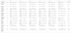
ResultsA total of 109 eyes from 55 subjects (aged 18–67 years; mean ± SD: 32.6 ± 12.6 years) were evaluated. Measurements were taken at 09:06 ± 19 min, 11:27 ± 17 min, 13:58 ± 23 min, 16:27 ± 19 min, and 18:49 ± 14 min. The sample comprised 37 women and 18 men, including 55 right eyes (RE) and 54 left eyes (LE). Mean spherical equivalent was −1.32 ± 2.31 D (RE) and −1.19 ± 2.23 D (LE). Mean axial length (AL) was 23.90 ± 1.13 mm (RE) and 23.89 ± 1.13 mm (LE). Participants reported an average sleep duration of 7.14 ± 1.04 h the night before measurements, with a mean time of 2.08 ± 0.84 h between waking and the first measurement. Descriptive statistics for self-reported lifestyle habits are summarized in Table 2.
Distribution of daily habits in the study population.
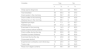
Mean values of each scleral parameter at all timepoints are summarized in Table 3, along with the corresponding CVs and p-values from inter-timepoint comparisons. No statistically significant differences were observed in scleral parameters across the day. Figs. 1 and 2 display violin plots illustrating the change from baseline at each timepoint (δ) for each scleral parameter.
Diurnal variation of scleral parameters.
Data are presented as mean ± standard deviation (SD) [range]. P-values correspond to the repeated-measures ANOVA (for normally distributed data) or the Friedman test (for non-normally distributed data) for comparisons across the five timepoints.
Abbreviations: CV= coefficient of variation; RE= right eye; LE= left eye.
 of sagittal height (minimum, maximum, astigmatic and mean), bulbar slope (minimum and maximum), and scleral best-fit sphere. Data are categorized into flattening and steepening trend groups. Violin plots show the distribution for the entire sample, while overlaid line plots display individual trajectories from a randomized subset of eight participants.")
Change from baseline at each timepoint (δ) of sagittal height (minimum, maximum, astigmatic and mean), bulbar slope (minimum and maximum), and scleral best-fit sphere. Data are categorized into flattening and steepening trend groups. Violin plots show the distribution for the entire sample, while overlaid line plots display individual trajectories from a randomized subset of eight participants.
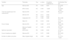
Statistically significant differences in the CVs of scleral parameters were observed for only a few lifestyle-related habits (Table 4). No significant correlations were found between scleral parameters and axial length (all p ≥ 0.263) or total screen time (all p ≥ 0.332). Additionally, no significant differences in scleral parameter CVs were found between individuals who did or did not engage in the following habits: morning water intake (p ≥ 0.333), smoking (p ≥ 0.076), morning physical activity (p ≥ 0.258), daytime physical activity (p ≥ 0.112), eye rubbing (p ≥ 0.090), and reading from non-digital media (p ≥ 0.096).
Statistically significant correlations between scleral parameters, lifestyle habits, and demographic variables.
Abbreviations: BS= bulbar slope; BFS= best-fit sphere;SH= sagittal height; RE= right eye; LE= left eye.
However, some significant correlations were identified in scleral parameter CVs and specific lifestyle behaviors. These included: washing the face in the morning (minimum BS RE; p = 0.007), having breakfast (minimum SH RE and LE; p = 0.016 and p = 0.001, resp.), drinking coffee in the morning (mean SH RE and maximum SH; p = 0.011 and p = 0.040, resp.), drinking coffee during the day (mean SH RE and scleral BFS RE; p = 0.016 and p = 0.040, resp.), screen exposure before bedtime (mean SH RE p = 0.036), and experiencing a stressful event (maximum BS RE and mean SH RE; p = 0.031 and p = 0.034, resp.). In addition, statistically significant sex-related differences were observed in the RE for minimum BS (p = 0.020) and astigmatic SH (p = 0.042). Figs. 3 and 4 illustrate these associations.


This is the first reported study to analyze changes in scleral geometry throughout the day. Although some fluctuations were observed in scleral parameters, no statistically significant changes were detected in BS or SH across timepoints. The magnitude of variability observed was consistent with the intrasession repeatability limits of the imaging system.
Bandlitz et al.19 reported that the CSP module demonstrates good repeatability for mean SH, with a mean difference of −0.9 μm. In our study, variability for this parameter was slightly higher, showing mean CVs of 0.70 ± 0.39 % and 0.88 ± 0.69 % for the right and left eye, respectively. Yang et al.20 found CVs below 0.96 % for mean SH, below 3.65 % for mean BS, and below 29.95 % for astigmatic SH in healthy eyes. Our results support these findings, suggesting that the observed variability falls within the instrument’s repeatability limits.
In contrast, a study by Read et al. reported significant diurnal changes in scleral thickness, particularly in the temporal quadrant.21 They observed peak thickness upon awakening and minimum values around midday. Although our measurements did not reveal statistically significant diurnal changes in scleral morphology, a non-significant decrease in BS at 11:30 was noted. This trend is consistent with Read et al.’s21 observation of a corresponding decrease in corneal thickness at that time, suggesting a possible shared physiological mechanism.The apparent stability of scleral geometry throughout the day supports its reliability as a baseline for contact lens fitting. Our findings confirm that SH remains stable in the short term, with no significant diurnal changes that could impact scleral lens fitting. Consequently, any variation in lens fit or position during wear is more likely due to lens-induced effects, such as mechanical indentation or deformation, rather than inherent changes in scleral morphology. Macedo-de-Araujo et al.8 demonstrated that scleral lens wear can induce changes in SH and reduce tangent angles in the nasal region at 7.5 mm and 8.00 mm chord lengths. However, our results indicate that diurnal rhythms do not meaningfully influence these parameters in the absence of lens wear and, therefore, scleral lens fitting.
Some lifestyle factors showed weak but statistically significant correlations with variations in scleral parameters. These correlations were asymmetrical between eyes, raising the possibility that habitual sleeping position may influence in scleral dynamics. Previous studies have linked sleeping posture to keratoconus progression, intraocular pressure fluctuations, and upper eyelid laxity.22–24 Notably, many of the most pronounced ocular changes —such as shifts in central corneal thickness and intraocular pressure —occur shortly after awakening. It is worth noting that the first measurement in this study was obtained approximately two hours after awakening, which may have missed some early-morning fluctuations. Such short-term changes could partly contribute to the diurnal stability observed in scleral morphology. This pattern suggests that mechanical pressure from the eyelids during sleep could contribute to morning asymmetries in ocular shape. Thus, sleeping position may partially explain the observed interocular differences in scleral CVs reported in this study.Despite limited correlation strengths (r ≤ 0.378), certain statistically significant trends emerged: participants with shorter sleep duration exhibited greater variation in BS and SH, and those with longer intervals between awakening and the first measurement displayed increased diurnal variability in minimum, mean, and astigmatic SH. Additionally, sex-based differences were noted, with women showing greater variation in minimum BS and astigmatic SH compared to men. However, these findings should be interpreted with caution due to the sample size and variability across groups.
This study has several limitations. First, no measurements were collected immediately upon awakening or during nighttime, which may have excluded time points critical for characterizing full diurnal patterns. Second, although interocular asymmetries were observed, the potential role of sleep posture (e.g., ipsilateral vs. contralateral eye contact with the pillow) was not evaluated. Therefore, the influence of sleep position on interocular asymmetries remains untested, highlighting the need for future studies to consider this factor. Third, the study sample was limited to healthy eyes; future investigations should include individuals with ocular surface disorders or ectatic diseases to determine if similar stability is preserved. Finally, our analysis focused on parameters measured within a 15 mm diameter, which may have missed regional or quadrant-specific changes in scleral geometry.
This study provides the first evidence of diurnal stability in the corneoscleral profile of healthy eyes, with no clinically meaningful changes observed in SH or BS. Observed fluctuations remained within the established repeatability limits of the CSP module, confirming the temporal reliability of these measurements. While statistically significant associations with lifestyle habits were limited, preliminary trends suggested possible relationships between scleral variability and factors such as sleep duration, breakfast consumption, and timing of the first measurement. These findings underscore the need for further research into how behavioral and physiological rhythms may influence ocular surface geometry.
FundingThe author Laura Barberán-Bernardos was supported by the Conselleria d'Educació, Cultura, Universitats i Ocupació of the Generalitat Valenciana within the Program ACIF (Subvenciones para la contratación de personal investigador predoctoral), reference number CIACIF/2022/073, cofinanced by European Social Fund.
The authors have no proprietary or commercial interest in the medical devices that are involved in this manuscript.


