


To evaluate the possible change in the optics of the human eye after iris constriction.
MethodsOcular aberrations were measured under natural viewing conditions in 26 eyes. The measured eyes fixated on a dim target while the contralateral eye was either occluded (so the measured eye had a large pupil) or highly illuminated (so the measured eye had a small pupil). The measured eyes fixated to a dim target placed 0.5 D beyond the subject’s far point. Zernike values obtained in both situations were compared within the same pupil diameter corresponding to the one obtained under the high illumination condition.
ResultsSignificant variation in some aberration coefficients were found between the two illumination conditions. Specially, spherical aberration (SA) increased significantly after pupil miosis (P = .0017). The mean increase of SA measured was 0.018 microns, for a 3-mm pupil. Mean values of other ocular aberrations also vary significantly after pupil miosis (changes were larger than the standard deviation of the repeated measurements). A mean paraxial hyperopic shift of one third of diopter was found after iris constriction.
ConclusionIris constriction slightly modifies the optics of the eye. The small hyperopic shift of the best image plane after iris constriction may be explained by a change in the lens shape and/or position.
Evaluar el posible cambio en la óptica del ojo humano tras la constricción del iris.
MétodosSe midieron aberraciones ópticas en condiciones de visión naturales en 26 ojos. Los ojos evaluados se fijaron en un objetivo atenuado, mientras que el ojo contralateral estaba oscurecido (para que el ojo evaluado tuviera la pupila grande) o muy iluminado(para que el ojo evaluado tuviera la pupila pequeña). Los ojos examinados fijados en un objetivo atenuado se situaron 0,5 dioptrías más allá del punto lejano del sujeto. Los valores de Zernike obtenidos en ambas situaciones se compararon dentro del diámetro de pupila correspondiente al obtenido en la situación de alta iluminación.
ResultadosSe observaron variaciones significativas en algunos coeficientes de aberración entre las dos condiciones de iluminación. Concretamente, la aberración esférica (AE) aumentó de manera significativa tras la miosis de la pupila (P = 0,0017). El aumento medio de la AE medida fue de 0,018 micrómetros para una pupila de 3 mm. Las medias de otras aberraciones ópticas también variaron significativamente tras la miosis de la pupila (los cambios fueron mayores que la desviación estándar de las mediciones repetidas). Se observó una desviación hipermetrópica paraxial media de un tercio de dioptría después de la constricción del iris.
ConclusiónLa constricción del iris modifica ligeramente la óptica del ojo. La pequeña desviación hipermetrópica del mejor plano de imagen después de la constricción del iris puede explicarse por el cambio en la forma y/o posición del cristalino.
It is well know that pupil size affects the optical quality of the human eye. After pupil miosis there are some well studied changes such as reduction in high-order aberrations,1 increasing diffractions effects,2 increase depth-of-focus3,4 and reduction of the Stiles Crawford effect5 that modify the optical quality of the eye. However, it remains unclear if the physical constriction of the iris causes a change in the optics of the eye as opposed to simply having a smaller pupil.
As evident example of the effect of the iris on the eye optics, some animal species such us diving birds or sea terns, accommodate by squeezing the lens with the outer portion of the iris.6–11 These species used pupil miosis to modify the power of the first surface of the lens increasing its curvature. Levy and Sivak9 found that certain aquatic birds possess an exaggerated accommodative ability resulting in a drastic change in the curvature of the anterior lens surface. Hess6 proposed that contraction of the ciliary muscle in birds such as the cormorant improves contact between the iris and the anterior lens surface; i.e. rather than acting on the lens directly; the ciliary muscle facilitates the action of the iris sphincter muscle on the lens. Walls7 and Goodge8 also suggested that accommodative effects of great magnitude are brought about by the action of iris sphincter contraction of the lens. Levy and Sivak9 proposed that the role of the iris is somewhat more passive. Rather than actively deforming the lens, contraction of the iris sphincter results in the formation of a rigid disc with a central aperture, the pupil. They suggested that contraction of the ciliary muscle pushes the malleable lens against the iris disc and the central lens bulges through the pupil. Then, iris sphincter contraction alone will not affect lens shape.
We have to consider that there are high differences between the ocular structures of the avian and human eyes (especially taking into account that the iris is firmly attached to the peripheral anterior lens surface in avian eyes). However, iris constriction also produces subtle modifications on the: shape and/or center of the pupil; the first surface of the lens; axial position of the lens; and tilt and/or centration of the lens.
The present study represents an effort to study possible modifications of the optics of the eye evaluating the changes of the ocular wavefront aberrations with and without iris constriction.
Materials and methodsSubjectsTwenty six subjects were enrolled in this study (mean 29.9 ± 8.1 years). In all cases they were healthy eyes without history of ocular abnormality, spherical refractive error below 5D and astigmatism less than 2D. Mean spherical equivalent was −1.38 ± 2.49D, ranging from 1.00 to −6.00 D. All subjects had a best corrected distance visual acuity of 20/20 or better. The subjects were recruited from Murcia, Spain. Subjects gave written informed consent and internal review board approval was obtained. The tenets of the Declaration of Helsinki were followed in this research.
Experimental procedureOcular wavefront aberrations were recorded using the Irx3 Wavefront Aberrometer (Imagine Eyes, Orsay, France). This device is based on the Hartmann-Shack aberrometer technique previously introduced by Liang et al.12 It uses a near infrared light source of wavelength 780 nm and a 32 × 32 microlens array sensor with an acquisition time of 33 ms. The built-in fixation targets pictures a polychromatic drawing of a balloon at the end of a road. The aberrometer was calibrated for a previous experiment13 and repeated measurements were obtained which gave us relevant information about its performances. See references 13 and 14 for details of the precision of the I × r3 aberrometer.13,14
We performed the same series of experiments in both eyes of each subject in monocular natural conditions (without pharmacological dilation or cyclopegia). Subjects did not wear refractive correction during experimental runs, being refractive error compensated by means of the internal Badal system incorporated in the aberrometer. We measured ocular aberrations under two illumination conditions. We showed the stimulus 0.5D beyond its far point being repeated for low (50 cd/m2) and high (3,000 cd/m2) illumination conditions. The measured eye fixated to the target while the counterpart eye was either occluded (showing the measured eye a large pupil) or highly illuminated with a white lamp (showing the measured eye a small pupil). The subject was instructed to maintain the object as clear as possible and the light used in the contralateral eye was indirect.
Prior to recording data, we ran at least one training trial in order to train the subject who was asked to keep looking at the smallest visible detail in the stimulus target. After checking that the task was well understood, we repeated the same procedure and recorded the resulting wavefront data in the form of Zernike expansions. The subject was allowed to blink during the procedure to avoid tear film aberration introduced by the air-tear film interface changes with time after a blink.15–17 We recorded 3 measurements for each pupil conditions.
Data analysisAll Zernike expansions were computed at least up to the 4th order for both light conditions (round pupil in all cases). We compared the values obtained for the smallest pupil diameter with those obtained under low lighting condition but computing them for the same pupil diameter used for the high lighting condition. Then, we compared both conditions with the same pupil diameter. In both cases we analyzed approximately the same Hartmann-Shack points (n > 110). Pupil center was chosen automatically using a pupil tracker incorporated in the aberrometer software.
Statistical analysis was performed using the SPSS software package (SPSS Inc, Version 11.5.1, Chicago, IL). A paired t-test was performed between Zernike coefficients obtained in both conditions (large and small pupil) in the same eye and calculated within the same pupil diameter (small pupil diameter). Differences between pupil diameters were also assessed with a paired t-test. Changes of Zernike coefficients after iris constriction where evaluated with a series of Bland-Altman plots. P values lower than 0.01 were considered statistically significant different.
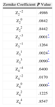
ResultsFigure 1 shows each individual Zernike term between values obtained for the high and low lighting conditions. Although the changes of aberrations depend on the subject and were small due to the small pupil diameter, we have noted significant variation in some aberration coefficients between both illumination conditions. To evaluate these changes figure 2 was created. This figure shows a series of Bland-Altman plots to assess agreement between low- and high-order aberrations computed for low and high lighting conditions. The figure is composed by several graphs showing the specific change for defocus, astigmatism, coma and spherical aberration. Table 1 shows the paired t-test values obtained for each Zernike coefficient between high and low lighting conditions. Pupil diameters measured for low and high lighting conditions were 6.45 ± 0.48 mm and 3.11 ± 0.42 mm, respectively. Reduction of pupil diameter between low and high lighting condition was 3.33 ± 0.53 mm (P < .01).
 between values obtained in each eye for the low lighting condition and those found for the high lighting condition. Error bars represent ± 1SD.")
. For each Zernike coefficient, X-axis represents the mean value between both lighting conditions, and Y-axis represents the differences between low and high lighting conditions.")
Paired t-test values obtained for each Zernike coefficient between real small pupil and simulated small pupil from large pupil condition
From figure 2 defocus analysis reveals that the maximum difference between both lighting conditions was 0.22 microns (corresponding to 0.68 D for a 3-mm pupil diameter). 83% of the eyes showed a large value of defocus under low lighting conditions (positives values). This represents a more myopic shift for a large pupil according to night myopia. The mean change was 0.05 microns corresponding to approximately one eighth of diopter. This difference was not significant (see Table 1). The mean value for both Z22 and Z3−1 were very similar for both lighting conditions. In contrast, our results revealed differences between mean values for Z2−2, Z31 and Z40. In particular, we found significant differences in horizontal coma (Z31, P = .004) as well as the both trefoils (Z33 and Z3−3). The only 4th-order coefficient with statistically significant differences was spherical aberration, SA (Z40, P < .01). There is a high percentage of set of measurements (88%, 46 out of 52) that shows an increase of the SA after iris constriction. The mean increase for 3-mm pupil was 0.015 mm. Although this variation is small we should consider that it has been computed for approximately a 3 mm pupil. This would correspond to 0.14 #um for a 5 mm pupil. That is higher than the mean SA of the eye18. The variation of SA found corresponds to a change of the best focal plane about 0.18 D of myopia.
From the aberration values of the patients studied (defocus and SA), we may obtain the spherical equivalent (SE)19 for each illumination level and in the situation of the dim illumination and large pupil to simulate the SE of the eye considering only the effect of the pupil (eye entrance pupil, getting smaller) without the effect of the iris. Then, we may compare the SE obtained with real small pupil (high illumination) with simulated small pupil (dim illumination). This difference will show us if there is a real refractive change produced by iris constriction. Using the metric based on the best fit of the wavefront to a sphere (also known as paraxial refraction),19 it can be obtained the SE by using the following equation:
being, r the pupil radius and Z the corresponding Zernike coefficients. We have obtained a significant (P = .00016) increase of the SE from the low light to the bright light condition, being the mean increase of 0.34 ± 0.39 D. Mean hyperopic shift was found for either myopic (18) or hyperopic (8) eyes. Then, under this metric, iris constriction generates a power reduction in the eye reflected by a hyperopic refraction shift about a third of diopter.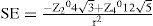
Paraxial refraction does not take into account the possible refractive changes generated by spherical aberration.20 Our results shows a decrease on Z40 after iris constriction (Figure 1 and 2) and an extra small myopic displacement of the best image plane could be expected when the metric used includes the effect of fourth-order spherical aberration (for instance for Zernike refraction). However, even when this effects are taking into account (i.e. under Zernike refraction), an hyperopic shift was also found since Z20 increase in mean when iris passes from a small radius to a large one (Figure 2). However, as mentioned before, that shift is small (around 1/8 of diopter) and non significant.
DiscussionWe have found statistically significant differences in several Zernike coefficients, being the SA change the most important. However, there are small changes in other coefficients which are not significant but the increase of the sample may lead a significant difference. We may consider several sources for these changes. Any physiological source to explain these changes should not have a random variation between eyes. Therefore, possible changes between eyes, eye position or gaze, tear film, subject respiration or microfluctuactions of the accommodation should not explain the differences found. In the next paragraphs we analyze the three non-random sources that could explain the outcomes reported.
Change in Accommodation by Depth-of-FocusSince the accommodation has not been paralyzed in our experiment, the accommodative state of the eye (so its optics) could differ between both lighting conditions. If we consider that when the pupil gets smaller the eye increases the depth-of-focus,3,4 then, we may expect a myopic shift after iris constriction causes by an involuntary accommodation. However, our results indicate the opposite. In addition, the increase of SA found when the pupil gets smaller implies that the eye has not accommodated since SA decreases during accommodation.13,21,22 Then, the increase of depth-of-focus should not play a role in the variations reported in this study.
Change in Pupil CenterWhen the pupil gets smaller a decentering of the pupil center may occur so the optics of eye change because we are analyzing another optical zone. As an example of the possible translation, we obtained several images of the patient's pupil. In these images we analyzed the variation of the pupil center in relation to the center of the circle of the first Purkinje image formed by two infrared diodes of illumination that don’t vary with pupil miosis. Figure 3 shows the position and the distance of the pupil center to the first Purkinje image of the diodes under low (a) and high (b) lighting conditions. The variation of the pupil center is systematically 0.14 mm nasal in average. This value agrees with normal pupil displacement reported in the literature (about 0.1 mm23).
 and high (b) lighting conditions. Pupil decentering values are included for both conditions for comparison.")
When the pupil is decentered in an eye, new Zernike values of lower order are generated being proportional to the decentration.24 For instance, a 6th-order aberration would generate a 4th-order aberration after a pupil translation.24 Considering this displacement, the eye should have a 6th-order SA of 0.057 μm in order to generate an increment of the 4th-order SA similar to that obtained (0.015 mm for 3-mm of pupil diameter).24 However, none of the eyes measured showed this quantity of 6th-order SA (all were smaller than this value). Then, our results cannot be supported by changes in the pupil center.
Change of Lens Shape and PositionThe last possibility is that the change may come from shape and position changes of the lens when the iris is constricted. This may be explained considering a direct relationship between the pupil edge and the first surface of the lens or an increase of the aqueous humor pressure. These small changes in the optics of the eye may be caused by small modifications of the lens shape. One may expect that a pressure on the pupil edge when the iris is constricted provokes a slightly convex curvature that increases the SA. If this pressure is not symmetric on the lens, asymmetrical aberration of 3rd-order may be generated.
Iris constriction could also modify lens position. In the case of the lens is moved towards the retina a decrease in the power of the eye is achieved (similar to the mechanism of accommodative intraocular lenses but in the opposite direction). This would explain the hyperopic shift obtained in our experiment. Furthermore, this explanation of our results will agree with previous literature25–27 indicating that night myopia (the eye becomes more myopic for a large pupil) is based on a change of the refraction state. In these studies, authors didn’t find a real change in the refractive status of the eye using artificial pupils, with the accommodation paralyzed. This indicates us that the myopic shift is not produced by a change in the pupil size, then, iris constriction is actively contributing to this shift.
In conclusion, our results shows a mean significant paraxial hyperopic shift of one third of diopter in the equivalent sphere after iris constriction that could be explained by a subtle change in the shape or a location of the lens, or probably combining both. Futures studies should include direct measurements of the lens's shape and location with accommodation. New anterior segment image techniques such as Scheimplug cameras28 would help to clarify the role of the iris in the modification of the eye optics.
This work has been supported in part by two grants from the Fundación SENECA, Comunidad Autónoma de la Región de Murcia (PI-42/00775/FS/01 and 00702//PPC//04, López-Gil, Programa SENECA 2007-Ayuda Estancias Investigadores Visitantes (Montés-Micó), and two grants from the Ministerio de Ciencia e Innovación Research Grants (#SAF2008-01114-E/# and #SAF2009-13342-E#, Montés-Micó).


