

This is to determine the prevalence of bacterial and parasitic flora in asymptomatic disposable and extended contact lens wearers in Lagos State, Nigeria.
MethodsThe study was carried out on 156 contact lenses from 78 asymptomatic wearers between the ages of 12 to 38 years. Eighty two disposable daily wear (DWCL) and 74 extended wear (EWCL) contact lenses were examined. The contact lenses’ swabs and the trays were transferred to the laboratory for microbial examination.
ResultsFifty two (70.27 %) extended wear contact lenses and 50 (60.98 %) disposable contact lenses examined were contaminated. Streptococcus spp. (4.23 %) were found in extended contact lenses and (3.9 %) in disposable contact lenses. Escherichia coli (15.49 %) were more in the extended contact lenses and were in higher proportion (14.74 %) than the other microorganisms in all the contact lenses. Klebsiella spp. (12.99 %) were more in the disposable lenses than in the extended wear lenses (12.69 %). there were more disposable lenses (41.56 %) with ‘no growth’. Amoebae were isolated from (6.49 %) disposable and (4.23 %) extended wear contact lenses. Seventeen (32.69 %) DWCL had mixed flora. There were significant differences between disposable and extended contact lenses, p<0.05.
ConclusionsEWCL present more microorganisms and pose threat to the users. DWCL had more amoebae, which calls for suitable lens care methods. Further studies may be needed to determine the level of care required for contact lens users in developing countries.
determinar la prevalencia de la flora bacteriana y parasitaria en usuarios asintomáticos de lentes de contacto desechables y de uso prolongado en el Estado de Lagos (Nigeria).
Métodosse estudiaron 156 lentes de contacto de 78 usuarios asintomáticos con edades comprendidas entre los 12 y los 38 años. Se examinaron 82 lentes de contacto de uso diario (LCUD) y 74 lentes de contacto de uso prolongado (LCUP). Los hisopos y los recipientes de las lentes de contacto se enviaron al laboratorio para realizar un análisis microbiano.
Resultadosde las lentes de contacto examinadas, 52 (70,27 %) de uso prolongado y 50 (60,98 %) desechables estaban contaminadas. Se encontró Streptococcus spp. en lentes de contacto de uso prolongado (4,23 %) y en lentes de contacto desechables (3,9 %). Se encontró más Escherichia coli en las lentes de contacto de uso prolongado (15,49 %) y en una proporción mayor (14,74 %) en comparación con el resto de microorganismos de todas las lentes de contacto. Se encontró más Klebsiella spp. en las lentes desechables (12,99 %) que en las lentes de uso prolongado (12,69 %). Hubo más lentes desechables sin crecimiento (41,56 %). Se aislaron amebas en las lentes de contacto desechables (6,49 %) y en las de uso prolongado (4,23 %). Diecisiete LCUD (32,69 %) presentaron flora mixta. Hubo diferencias significativas entre las lentes de contacto desechables y las de uso prolongado, p<0,05.
Conclusioneslas LCUP presentan más microorganismos y suponen un mayor riesgo para los usuarios. Las LCUD presentaron más amebas, por lo que es necesario aplicar métodos adecuados para el cuidado de las lentes. Es posible que hagan falta más estudios para determinar el nivel de cuidado necesario para los usuarios de lentes de contacto en los países en vías de desarrollo.
The inconvenience of wearing the conventional spectacles had led to the development of plastic corrective contact lenses (CL) worn directly over the cornea to improve vision. The use of contact lenses (CL) had increased remarkably because of its optical, occupational and cosmetic advantages. However, Devonshire et al1 reported that the problem in contact lens wear was the presence of bacteria and other microorganisms; because some contact lens wearers had developed microbial keratitis. Martins et al2 observed the presence of fungi, parasites and bacteria in contact lens swabs cultures. It has been reported that the environment, the type of contact lens, the duration of wear, and the type of CL cleansing solution determined the microbial load on the contact lenses.3–5Staphylococcus epidermidis, Staphylococcus aureus, Enterobacter and Pseudomonas species found in healthy eyes, were also observed on soft contact lenses of healthy persons.6Acanthamoeba species were also found in contact lenses. These amoebae lived in the bio-films of other organisms and decaying organic materials, where they feed on bacteria and other microscopic organisms.
Acanthamoebae thrive well in water supply systems and proliferate on the inside surfaces of pipes. They were also found in moist soil and mud. These amoebae could be resistant to dry environments, chlorine, and many contact lenses cleansing antiseptics. They are capable of feeding on living tissues. If found in human corneal tissues could cause disease and this has been related to CL wear. Therefore, CL wearers using tap water to clean their contact lenses instead of the prescribed solution, might face the risk of being infected by these parasites, which could cause resistant eye infections. This could endanger the cornea. We often learn about the destructive effects of these parasites on the cornea in the laboratory, at the time it was too late for any useful remedy. Thus there should be continuous monitoring of these emerging pathogens, which could cause serious eye infections.7
Many authors had reported the presence of microbial organisms on contact lenses of Caucasians wearers, but none on black contact lenses wearers.8–10 The reason may not be farfetched, because contact lens wear is relatively new to Africa. Contact lens wearers have increased in Nigeria, where the climatic conditions and the environment favour the growth of microorganisms. There may be more problems associated with contact lens wear in the developing nations than in the industrialized nations. Lagos is a typical city in a developing nation in Africa. It is an industrial city and a densely populated area of Nigeria. It is the former capital of Nigeria. The inhabitants are exposed to dirty environment, water and soil where these microorganisms comfortably thrive. Therefore, it is important to identify the bacteria and the parasites that contaminate contact lenses (CL) in Lagos (Nigeria) and to determine if the microbial contaminants are more in either extended wear (EWCL) or disposable daily wear contact lenses (DWCL).
MethodsThis study was carried out between August 2002 and December 2003 in private eye clinics in Lagos, because of the relatively high population density of contact lens wearers in Nigeria. The study population consisted of 78 asymptomatic contact lens wearers between the ages of 12 to 38 years. We were unable to find subjects above 40 years that wear contact lenses, because it has not obtained general acceptance. Contact lens (CL) wearers with eye infections or under any therapeutic or diagnostic eye drops were excluded from the study. The disposable daily wear (DWCL) and extended wear (EWCL) contact lenses included in the study must have been worn for 4 hours and above. The subjects were the regular contact lens patients that visited the clinics for their lens care regimen. The study was carried out after oral and written consent of the subjects were obtained. The study was conducted in accordance with the tenants of the declaration of Helsinki. During this period, 156 soft hydrogel contact lenses were examined for the presence of microbial and parasitic organisms; there were 82 (52.6 %) daily disposable soft hydrogel contact lenses (DWCL) and 74 (47.4 %) extended wear silicone hydrogel (S-H) contact lenses (EWCL). The (DWCL) lenses were Acuvue 2 (Etafilcon A, 58 % water content, ionic) manufactured by Johnson and Johnson (Jacksonville, FL) and Soflens 38 (Polymacon, 38 % water content non-ionic) manufactured by Bausch and Lomb (Rochester, NY). All the extended wear lenses were made of S-H material from Bausch and Lomb (PureVision, Balafilcon A, 36 % water content, ionic surface treated lens). Sterile cotton-tip swabs moistened with sterile saline were used in the collection of samples from the contact lenses in the sterile contact lens tray. The participants were asked to place their contact lenses on the sterile trays after the washing of their hands with sterile water and soap. Sterile swabs were rotated on the contact lens surface immediately they were placed on the sterile tray. Each contact lens examined was washed with three drops of sterile saline into a lens tray. Each participant was afterwards asked to wash their contact lenses (CL) with a contact lens disinfecting solution before reinsertion on the cornea. The swabs and the contact lens trays were thereafter sent to the laboratory for examination.
Contact lens bacteriologic studyFour swabs were collected from each subject's contact lenses, two from each eye contact lens in the trays. The swabs and the trays were sent to the laboratory before 12 hours for culture and microscopy. Each swab was used to inoculate two culture media, the first was used to streak a blood agar, chocolate agar and MacConkey agar plates; the second was used to streak a Sabouraud dextrose agar plate. Gram staining was carried out in each case. The tip of each swab was broken off (2 to 4cm above the tip) and placed into a meat broth. All plates and broths were incubated at 37°C to allow for bacterial growth and held for 48 hours to ascertain either “growth” or “No growth” and all organisms found were identified. The Sabouraud's dextrose agar plate and the meat broth were held for 1 week and the organisms present were identified. The colony counts on the plates were recorded.
Positive cultureThe organisms that grew on any of the media were identified. Staphylococcus aureus, coagulase negative Staphylococcus, and Streptococcal species isolated only in meat broth were considered to be contaminants. “Positive” cultures fulfilled one of the following criteria: the organisms isolated on at least one solid medium, organisms isolated from two or more media, gram negative or anaerobic organisms isolated only from the meat broth. Positive cultures were divided into two categories. The first, were the normal conjunctival flora, such as coagulase-negative Staphylococci. Although these organisms could be opportunistically pathogenic, they frequently colonize the ocular surfaces of normal subjects. Secondly, were the potential pathogens that consisted of any microorganisms other than coagulase negative Staphlococci.11
Parasitological cultureNon nutrient agar (NNA) was used with an overlay of Escherichia coli, for the growth of Acanthamoeba spp and other Amoebae. The specimen from each CL tray was simply introduced into the surface of the plate without streaking or breaking the surface. Two plates were inoculated for incubation at 25 and 37°C because some species might not grow at higher temperature. The plates were examined for trophozoites and cysts directly under the microscope. Trophozoites were observed in 24 to 48 hours. They moved and covered the entire plate surface and on further incubation some turned into cysts. The plates were observed for at least 10 days.
Statistical analysisSPSS statistical software was used for data analysis. Mann-Whitney U-test statistical ranking for unpaired observations was employed in the determination of the significant differences between the contaminated disposable and the extended wear contact lenses, p<0.05 was considered statistically significant.
ResultsOut of the 156 contact lenses examined 54 (34.62 %) showed ‘No growth’. Contact lenses with amoebae were 8 (5.13 %), while bacteria contaminated 94 (60.26 %) contact lenses (Table 1). A total of 35 (22.44 %) of the contact lenses had mixed flora. It was observed that 35 of the contact lenses showed pathogenic organisms. Staphylococcus epidermidis was found in 6 (3.85 %) contact lenses. Escherichia coli in 22 (14.74 %) contact lenses, Klebsiella spp. in 20 (12.82 %) contact lenses and Staphylococcus aureus in 7(4.49 %) contact lenses were observed. Other bacterial isolates such as Pseudomonas spp. and Corynebacterium spp. were found in small number of 5 (3.2 %) and 2 (1.3 %) contact lenses, respectively. Out of the 74 EWCL, 52 (70.27 %) were contaminated, while with the 82 disposable contact lenses examined, 50 (60.98 %) were contaminated. Amoebae isolated from the disposable lenses were more than that observed with the extended wear contact lenses.
Table 2 revealed that many of the participants wore daily disposable contact lenses (DWCL) and 17 (32.69 %) of the DWCL swabs had mixed flora. Klebsiella spp. 10 (12.99 %) were found in daily disposable contact lenses (DWCL), while Escherichia coli was more in the extended S-H contact lens (EWCL). Thirteen EWCL had mixed flora (Table 3). Mann-Whitney U-test revealed significant differences between disposable and EWCL with microbial isolates (calculated U1=−1524.9 and critical U=526), p<0.05.
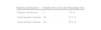
Organisms on 74 disposable contact lenses (DWCL)
| Isolated organisms | No. of swabs | Percentage (%)* |
| Streptococcus spp. | 3 | 3.9 |
| Escherichia coli | 11 | 14.29 |
| Pseudomonas spp. | 4 | 5.19 |
| Klebsiella spp. | 10 | 12.99 |
| No growth | 32 | 41.56 |
| Klebsiella ssp. + Pseudomonas spp. | 2 | 2.6 |
| Streptococcus spp. + Staphylococcus | 5 | 6.49 |
| Staphylococcus ssp. + Klebsiella spp. | 2 | 2.6 |
| Klebsiella spp. + Escherichia coli | 1 | 1.3 |
| Klebsiella spp. + Streptococcus spp. | 2 | 2.6 |
| Klebsiella spp. + S. aureus + Escherichia coli | 3 | 3.9 |
| Escherichia coli + Staphylococcus aureus | 2 | 2.6 |
| Amoebae sp. | 5 | 6.49 |
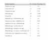
Organisms on 82 extended wear contact lenses (EWCL)
| Organisms | No. of swabs | Percentage (%)* |
| Streptococcus spp. | 3 | 4.23 |
| Staphylococcus epidermidis | 6 | 8.45 |
| Staphylococcus aureus | 7 | 9.86 |
| Escherichia coli | 11 | 15.49 |
| Pseudomonas spp. | 1 | 1.41 |
| Corynebacterium spp. | 2 | 2.81 |
| Klebsiella spp. | 9 | 12.68 |
| No growth | 22 | 30.99 |
| Streptococcus spp. + Staphylococcus | 6 | 8.45 |
| Escherichia coli + Streptococcus spp. | 2 | 2.81 |
| Staphylococcus spp. + Klebsiella spp. | 1 | 1.42 |
| Escherichia coli + Staphylococcus aureus | 1 | 1.42 |
| Amoebae sp. | 3 | 4.23 |
It was found that 52 (70.27 %) EWCL and 50 (60.98 %) DWCL examined were contaminated. Streptococcus spp. were found in EWCL (4.23 %) and in DWCL (3.9 %). Escherichia coli (15.49 %) was more in the EWCL and was found in higher proportion (14.74 %) than the other microorganisms in the contaminated contact lenses. Klebsiella spp. (12.99 %) were more in the DWCL than in the EWCL (12.69 %). However, there were more DWCL with ‘no growth’ (41.56 %). Amoebae isolated from the DWCL (6.49 %) were more than that observed with the EWCL (4.23 %). Seventeen (32.69 %) of the DWCL had mixed flora. There were significant differences between the microbial contents of the DWCL and that of the EWCL (calculated U1=−1524.9 and critical U=526), p<0.005.
DiscussionThere is a continuous increase in the use of contact lenses in Nigeria because of the optical, occupational and cosmetic advantages to individuals. Several authors reported that the introduction of contact lenses was associated with increase in ocular microbial complications.1,12 The unique structure of the human eye, the use of contact lenses and the constant exposure of the eye directly to the environment renders it vulnerable to a number of uncommon infectious diseases caused by parasites, and bacteria. Some of these infectious eye diseases, prior to the invention of contact lenses, were rare. Thus new opportunities were offered to these microorganisms when people started wearing contact lenses. Host defenses directed against these pathogenic microorganisms, once anatomical barriers were breached, were usually inadequate to prevent loss of vision.6,9 Therefore, necessary precautions are required to protect the eye from these opportunistic organisms. These microorganisms and their pathogenic effects might be different from country to country, particularly in the developing countries.13–16 Therefore, the timely identification of the microorganisms found in contact lenses of African wearers is of paramount importance.
Many of the participants in the study (72 contact lens wearers) were young adults, below the age of 40 years and others were children. This revealed that young adults were more adventurous in trying out new visual aid gadgets. The parasitic and bacterial flora found in these contact lenses of asymptomatic wearers might be from the environment, water, physical contact, or from unhygienic habits of the wearers. Therefore timely treatment of corneal abrasion as a result of contact lens wear is important. Contact lens users with mild ocular surface diseases or corneal abrasion might be at risk of microbial keratitis.17,18
The Staphylococcus epidermidis, Staphylococcus aureus, Escherichia coli (E. coli), and Klebsiella spp. were the most common microorganisms found in this study. The study carried out in UK revealed that Staphylococcus epidermidis was found to be more in normal conjunctival flora.19 Other similar reports had confirmed the study, with slight variations in the percentages of occurrences.20–22 In this study, E. coli was found in higher percentage than other microorganisms (Tables 2 and 3) on the contact lenses. Larkin and Leeming23 studied normal ocular flora and compared it with that of the asymptomatic contact lens users and found that Staphylococcus epidermidis was more amongst the non contact lens wearers. Sankaridurg et al.6 found that ocular microorganisms were lesser in asymptomatic contact lens users than during corneal infiltration. They6,24 observed some differences in the microbial load between non contact lens wearers and asymptomatic contact lens wearers. This could be a major reason of the low percentage of Staphylococcus epidermidis observed in this study.
The presence of E. coli in the examined contact lenses could be from the use of contaminated water. Free living amoebae had been isolated from the dust, contact lenses, domestic water and swimming pool.24–26 Kamel and Norazah27 reported the first case of Acanthamoeba keratitis in a female's contact lens. It has been suggested that bacteria found in eyelids, conjunctiva and tear film might have had a contributory role in the pathogenesis of Acanthamoeba keratitis.28 It has also been reported that Acanthamoeba keratitis occurred more in contact lens wearers, probably as a result of the contaminated tap water used for the lens care. There were further evidences that soft contact lens wearers could be at greater risk for protozoan infection.29,30 The study agreed with this observation that daily disposable contact lenses (soft contact lenses) were more predisposed to the amoebea31 (Table 2).
It was also observed in this study that there were significant differences in the microbial presence between the DWCL and the EWCL. The observation in this study agreed with the clinical trials report of Fonn et al,32 which revealed that the EWCL yielded much higher bacterial adverse response rate than the DWCL. However, Gopinathan et al.10 reported that the increase in the length of lens wear did not result to the predictability increase in the bacteria colonization of the contact lenses. Those authors argued also that the bacterial types present in the normal ocular microbiota were rarely associated with diseases. Therefore the EWCL did not alter the frequency of bacterial colonization of lenses in neophyte wearers in both Australians and Indians.10
It is therefore obvious that there are controversies about the effect of soft contact lenses on ocular microbiota and the associated diseases. Many authors reported that asymptomatic lens wear for extended periods did increase ocular microbiota23,33,34 and others reported that asymptomatic lens wear for extended periods did not increase normal ocular microbiota.10,35–37 However, Efron et al38 suggested that ocular diseases of contact lens wearer could be as a result of noncompliance or omission of surfactant cleaning rub and rinse steps, the use of disinfecting solution of marginal efficacy and lenses that attract and rapidly deposit protein. Thus, the lens care regimen is an important factor for consideration on subjects that showed ‘no growth’ among the daily and extended contact lenses wearers.
A total of 54 (34.62 %) contact lenses examined had ‘No growth’ and 35 (22.44 %) of the contact lenses had mixed flora. It was observed that 35 (22.44 %) of all the contact lenses had pathogenic organisms, 8 (5.13 %) parasitic infested contact lenses and 94 (60.26 %) bacterial contaminated contact lenses in healthy eyes. Staphylococcus epidermidis, a normal ocular flora, was found in 6 (3.85 %) contact lenses. Klebsiella spp. and Staphylococcus aureus were found in 20 (12.82 %) and 7 (4.49 %) contact lenses, respectively.
This study attempts to suggest that daily disposable contact lenses (CL) should be more suitable for wearers in Nigeria and it gives a clue that more care is required for the use EWCL in Nigeria. However, further studies may be required to determine the appropriate care methods suitable for soft contact lens wearers in Nigeria, in order to reduce the degree of parasitic contamination.
Conflict of interests
Authors declare that they don’t have any conflict of interests.


