

The aim of the study was to evaluate the role of anti-VEGF therapy as an adjunct to laser ablation therapy in severe aggressive posterior retinopaty of prematurity (AP-ROP).
MethodsMedical records of premature infants with a primary diagnosis of AP-ROP treated with 0.625mg/0.025ml intravitreal bevacizumab (IVB) in addition to standard laser photocoagulation as a salvage therapy or primarily combined with laser in a university clinic were reviewed, retrospectively. The anatomical results and complications were evaluated after treatment.
Results15 eyes of 9 patients with a mean gestational age of 28.3 weeks (range, 26–31w) and mean birth weight of 1090g (range, 860–1330g) were included in the study. They all had Zone 1 or posterior Zone 2 plus disease staging between severe 3 and 4a. Twelve eyes were treatment naive at the beginning. The mean follow-up was 19.5±11.8 months (range, 11–40 months). The disease regressed totally in 6 eyes (40%), stayed stable as stage 4a in 1 eye (6.7%), progressed to and stabilized at stage 4a in 3 eyes (20%) and progressed to stage 5 in 3 eyes (20%) within 7–10 days. Two eyes (13.3%) developed hypotony and cataract.
ConclusionThe association of IVB and laser ablation might decrease the progression rate in severe AP-ROP. Prompt regression of iris neovascularization encourages its use in cases with pupillary rigidity to allow for laser treatment. When used as a salvage therapy it may not change the overall result dramatically.
El objetivo del estudio fue la evaluación del papel de la terapia anti-VEGF como complemento a la terapia de ablación por láser en la retinopatía posterior agresiva severa del prematuro (AP-ROP).
MétodosSe revisaron retrospectivamente los informes médicos de niños prematuros con diagnóstico primario de AP-ROP, tratados con 0,625 mg/0,025ml de bevacizumab intravítreo (IVB), además de fotocoagulación con láser estándar como terapia de rescate, o previamente combinado con láser, en una clínica universitaria. Tras el tratamiento se evaluaron los resultados y complicaciones anatómicas.
ResultadosSe incluyeron en el estudio 15 ojos de 9 pacientes con una edad media de gestación de 28,3 semanas (rango, 26-31 semanas) y un peso medio al nacer de 1090g (rango, 860-1330g). Todos ellos tenían enfermedad de zona 1 o zona 2 plus posterior, con niveles comprendidos entre 3 severa y 4a. Al inicio, doce ojos no habían sido tratados anteriormente. El seguimiento medio fue de 19,5±11,8 meses (rango, 11-40 meses). La enfermedad mostró una regresión total en 6 ojos (40%), permaneció estable en estadio 4a en 1 ojo (6,7%), progresó y se estabilizó en estadio 4a en 3 ojos (20%) y progresó a estadio 5 en 3 ojos (20%), durante un periodo comprendido entre 7 y 10 días. Dos ojos (13,3%) desarrollaron hipotonía y cataratas.
ConclusiónLa asociación de IVB y la ablación por láser puede hacer disminuir la tasa de progresión en la AP-ROP severa. La rápida regresión de la neovascularización del iris estimula su uso en casos con rigidez de la pupila, dejando margen a la terapia con láser. Su uso como terapia de rescate puede no modificar drásticamente el resultado general.
Retinopaty of prematurity (ROP) is a proliferative disorder of the developing retina that continues to be a major cause of blindness of children in the developed and developing world.1,2 Both Cryo-ROP3 and Early Treatment ROP4,5 studies demonstrated improved structural and functional outcomes with peripheral ablation. Although incidence of retinopathy was similar in these two studies, an increase in the occurrence of Zone 1 ROP was noted in the Early Treatment ROP cohort. The increase in Zone 1 ROP is most likely due to increased survival of ultrapremature infants (gestational age<26 weeks, birth weight<750g, or both) because of improved neonatal care.
Aggressive posterior ROP (AP-ROP) is characterized by Zone 1 or posterior Zone 2 ROP requiring treatment per Early Treatment ROP standards (any plus disease or stage 3 disease). These infants are characterized by flat neovascularization (stage 3), plus disease, or both generally occurring by postmenstrual age of <34 weeks. Management is complicated by a poor view of the fundus because of persistent tunica vasculosa lentis and persistent fetal vasculature. Complete laser ablation may not be possible in a single session, and repeat laser ablation may be warranted. The prognosis of AP-ROP is still guarded even with treatment; the cryotherapy for Retinopathy of Prematurity Cooperative (Cryo-ROP) Study reported a 77.8% unfavorable outcome rate with cryotherapy, and the Early Treatment for Retinopathy of Prematurity Cooperative Group (ET-ROP) reported a 55.2% unfavorable outcome rate with laser photocoagulation in Zone 1 disease.3,4
It is currently understood that ROP is a biphasic disease consisting of an initial phase of oxygen-induced vascular obliteration followed by a period of hypoxia-induced vessel proliferation.6 Anti-VEGF therapy may be effective in the second phase and as a single dose because there is theoretically only 1 burst of VEGF.7 This is in contrast to other ocular neovascular conditions, such as age-related macular degeneration and diabetic retinopathy, where aberrant VEGF production often recurs as a result of longstanding disease. Sonmez and associates reported a significant increase in the intravitreal levels of VEGF in vascularly active stage 4 disease, indicating that the administration of bevacizumab should be before the onset of this increase, such as during stage 3.8
On the basis of reports of the successful use of anti-VEGF agents in the treatment of retinal proliferative diseases, several groups have conducted preliminary trials with intravitreal bevacizumab (IVB) (Avastin; Genentech, San Francisco, CA) in small series of patients with ROP as an adjunct (to laser) or salvage therapy or as monotherapy with favorable anatomical outcome in some of the cases.9–30
Currently, the results of BEAT-ROP study (Bevacizumab eliminates the angiogenic threat of retinopathy of prematurity) which is a randomized controlled Phase II multicenter trial exploring the safety and efficacy of IVB for Stage 3 ROP in severe Zone I or posterior Zone II in 150 premature neonates have been released. The babies were randomized to receive confluent laser or IVB 0.625mg as primary therapy in both eyes. The results showed that bevacizumab uniformly reduced neovascularization and plus disease within 48h. Both persistence/recurrence of vascular activity and progression to RD were significantly lower in the study group than in the laser-treated (control) group, especially in eyes with Zone I disease.25 After encouraging results of BEAT ROP study, in the present study, we have investigated the efficacy of intravitreal bevacizumab (IVB) treatment combined with laser ablation or as a salvage therapy after laser ablation in AP-ROP.
Patients and methodsMedical records of premature infants with a primary diagnosis of AP-ROP treated with intravitreal bevacizumab (IVB) in addition to standard laser photocoagulation as a salvage therapy or primarily in a university clinic were reviewed retrospectively. All of the surgical procedures had been performed by two surgeons (SO, GG), including IVB injections and laser applications after getting a consent form from the parents explaining the off-label use of bevacizumab and the potential risks of the drug and the injection.
Surgical techniqueEyes received a near confluent pattern of indirect diode laser photocoagulation to the avascular retina, immediately anterior to the border of the vascular zone extending to the ora serrata for 360° under general anesthesia.
Bevacizumab (0.625mg/0.025ml) was injected intravitreally using a 30-gauge needle placed 1mm behind the limbus, immediately after completion of photocoagulation or later as salvage therapy in those not responding to laser photocoagulation.
The data included the demographic features of the babies, associated systemic problems, general health conditions of the babies as well as pre and postoperative ophthalmologic features; pupillary dilation, iris neovascularization state, location (zone), extent (clock hours) and stage of the disease and presence of plus disease or vitreous hemorrhage. Treatment details including the type and amount of anti-VEGF used and the number of laser burns were noted. Patients were followed weekly during the 1st month and monthly thereafter if not requested otherwise. During the follow-up period, all of the above data were reviewed to determine the postoperative complication and anatomical success rate.
ResultsFifteen eyes of 9 consecutive patients (6 boys, 3 girls) were included in the study. Mean gestational age was 28.3 weeks (range, 26–31 weeks), mean birth weight was 1090g (range, 860–1330g) and mean follow up time was 19.5±11.8 months (range, 11–40 months). Seven of 9 infants had severe systemic problems (8 had respiratory distress syndrome, 2 had intraventricular hemorrhage and 3 had sepsis) at postpartum period. Demographic data are listed in Table 1. They were all referred from other hospitals to our center for ROP treatment.
Demographic data of the babies.
| Case | Gestational age (weeks) | Birth weight (g) | Age at treatment (weeks) | Systemic problems |
| Case 1 | 31 | 1000 | F | Twin, RDS |
| Case 2 | 28 | 1100 | M | RDS |
| Case 3 | 28 | 1100 | F | Blood transfusion, RDS |
| Case 4 | 31 | 1330 | F | Sepsis, RDS, IVH, Twin |
| Case 5 | 29 | 1250 | F | RDS |
| Case 6 | 26 | 860 | M | RDS |
| Case 7 | 26 | 900 | F | Sepsis, RDS |
| Case 8 | 28 | 1100 | M | Sepsis, RDS |
| Case 9 | 28 | 1180 | M | IVH |
RDS, respiratory distress syndrome; IVH, intraventricular hemorrhage; M, male; F, female.
All eyes were Zone 1 (13 eyes) or posterior Zone 2 (2 eyes). All eyes had plus disease before treatment (Table 2). They were all severe stage 3 or 4a eyes and 12 eyes were treatment naive at the beginning and two patients were sent from other hospitals to our center after inadequate laser treatment. Mean number of laser spots applied in our clinic was 2136±585 spots (range, 1900–3000). Two eyes (13%) received IVB injection at the same session with laser photocoagulation because of the presence of severe plus disease, and 13 eyes (87%) received IVB later as a salvage therapy.
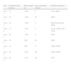
Clinical Characteristics of Patients.
| Case/eye | Zone | Stage | Plus disease pre/post-treat. | Pupil problem (RI or poor dilatation) pre/post-treat. | Vitreous hemorrhage pre/post-treat. | Follow-up (months) | Prognosis |
| Case 1 | |||||||
| R | Zone 1 | 3 | +/− | +/ | −/+ | 14 | Anterior segment ischemia |
| L | Zone 1 | 3 | +/− | +/ | −/+ | Progression (stage 5)b | |
| Case 2 | P.Zone 2 | 3 | +/− | −/− | −/− | 12 | Progression (stage 4a) |
| Case 3 | Zone 1 | 4a | +/− | −/− | −/− | 14 | Stabile (stage 4a) |
| Case 4 | |||||||
| R | Zone 1 | 4a | +/− | −/− | +/+ | 40 | Progression (stage 5)b |
| L | Zone 1 | 4a | +/− | −/− | −/− | Regression | |
| Case 5 | P.Zone 2 | 4a | +/− | −/− | −/− | 39 | Regression |
| Case 6a | |||||||
| R | Zone 1 | 3 | +/− | +/− | +/+ | 11 | Anterior segment ischemia |
| L | Zone 1 | 3 | +/− | +/− | −/− | Regression | |
| Case 7 | |||||||
| R | Zone 1 | 3 | +/− | −/− | −/+ | 23 | Progression (stage 5)b |
| L | Zone 1 | 3 | +/− | −/− | +/− | Regression | |
| Case 8 | |||||||
| R | Zone 1 | 3 | +/− | +/− | −/− | 12 | Regression |
| L | Zone 1 | 3 | +/− | +/− | −/− | Progression (stage 4a) | |
| Case 9 | |||||||
| R | Zone 1 | 3 | +/− | +/− | +/− | 11 | Progression (stage 4a) |
| L | Zone 1 | 3 | +/− | +/− | −/− | Regression | |
All of the eyes had a significant decrease in plus disease as observed by first clinical examination in the 1st week. Eight eyes of 4 infants (cases 1, 6, 8 and 9) had pupillary problems (anterior neovascularization and poor dilation) before treatment, and developed beneficial response to treatment with decreased engorged iris vessels permitting dilation of the pupils. The disease regressed totally in 6 eyes (40%), stayed stable as stage 4a in 1 eye (6.7%), progressed to and stabilized at stage 4a in 3 eyes (20%) and progressed to stage 5 in 5 eyes (33.3%) within 7–10 days. Two of the 3 eyes with progression to stage 5 (case 1 and 6) developed hypotony and cataract. Macula was on in 66.6% of the eyes at the last control visits. Of the 4 eyes having mild vitreous hemorrhage before treatment, only one regressed after treatment (case 7/L) and other three eyes progressed to stage 4a or 5 (Table 2). New vitreous hemorrhage developed in another 3 eyes following treatment all of which progressed to stage 5.
No alteration in blood pressure or systemic condition was observed suggesting the absence of systemic toxicity of the bevacizumab in any of the patients following treatment.
DiscussionAP-ROP is an uncommon disease and comprises 10% of ROP requiring treatment. It is well established that Zone 1 disease often progresses despite conventional therapies; unfavorable outcomes up to 55–78% such as macular fold and dragging, retrolental mass and retinal detachment could be observed in APROP eyes as proven by prospective controlled studies.26 The disappointing results in APROP stimulated many authors to find better treatment modalities for these eyes. The use of bevacizumab in ROP has become a relatively new topic in retinal research and the question still remains in the adequacy of the use of bevacizumab for the treatment of ROP.
Currently, one of the two randomized controlled studies exploring the safety and efficacy of IVB for ROP is named as “Pan-VEGF blockade for the treatment of retinopathy of prematurity (BLOCK-ROP), clinical trials identifier NCT00702819” which is a Phase I multicenter study of the safety and efficacy of unilateral IVB in premature neonates with posterior ROP that is unresponsive to laser treatment. However, this phase of BLOCK-ROP was terminated due to insufficient enrollment—ROP progression with timely and effective laser for APROP was rare at BLOCK-ROP study centers. Two infants were enrolled over the course of a year (unpublished data).31 A second phase of the BLOCK-ROP study (NCT01232777) targets all treatment-eligible infants (Type-1 threshold ROP), with an internal control for more rigorous comparison of treatment efficacy. This dose-ranging Phase II study will aim to demonstrate noninferiority of IVB compared with standard-of-care laser. In this randomized three-armed trial, one group of infants will receive 0.75mg IVB in one eye and laser treatment in the fellow eye; a second group will receive a lower dose of 0.625mg IVB in one eye and laser treatment in the fellow eye; and a third group of infants will receive laser photocoagulation in both eyes. It is hoped that, by this study design, the relative risk of increased myopia, amblyopia and functional outcomes can be assessed, since the eye treated with IVB can be compared with the fellow laser-treated eye of the same infant. Additionally, a lower effective dose of bevacizumab may be established.
The second study, “Bevacizumab eliminates the angiogenic threat of retinopathy of prematurity (BEAT-ROP),” is a Phase II multicenter trial in which 150 premature neonates with severe stage 3 ROP in Zone I or posterior Zone II were randomized to receive confluent laser or IVB 0.625mg as primary therapy in both eyes. The results showed that out of 143 infants who survived to week 54 postmenstrual age, ROP recurred in 4% of the bevacizumab group and 22% of the laser group.25 Both persistence/recurrence of vascular activity and progression to RD were significantly lower in the study group than in the laser-treated (control) group, especially in eyes with Zone I disease. Still there is a considerable concern regarding the choice of anti-VEGF, its dosage, timing of injections, the possible need for multiple injections, the reality that local complications of ocular trauma or endophthalmitis may occur, and, most importantly, the possibility that both acute and long-term systemic complications may be caused by anti-VEGF therapy since the BEAT-ROP study was not powered to assess safety. Shah et al. described visual acuity and electroretinography findings 3½ years after the first IVB injection in a case of APROP. They suggested that IVB did not have an adverse effect on the ERG but could have on the final visual acuity.32
An important advantage of IVB therapy is the continuation of peripheral retinal vascularization after treatment. Hoang et al. evaluated fluorescein angiography of recurrent ROP after initial IVB treatment. After 2 months of IVB injection they documented circumferential anastomosis at the bevacizumab induced regression site with radial vessels progressing anteriorly to form a second stage 3 complex. This case also provided further support that IVB did not necessarily inhibit subsequent retinal vascular development.33
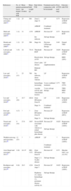
There are several articles reporting the use of IVB for treatment of Zone 1 and posterior Zone 2 diseases, as monotherapy or in combination with conventional laser therapy or vitrectomy9–30 (Tables 3 and 4). There are 109 eyes of 63 patients treated with IVB in conjunction to lasers all of which had APROP (Table 3). Each report was of a different design and used a different dose of bevacizumab, the most commonly used dose being 0.625mg. Of the 109 eyes in the studies, 41 (38%) eyes received IVB as an adjunct to laser photocoagulation initially, 35 (32%) eyes received IVB as a salvage therapy for severe laser-refractory ROP and 33 (30%) eyes received IVB for the limited visibility of the fundus because of vitreous haze or hemorrhage and other media opacities and poor pupillary dilatation or in infants who are too unstable medically to safely undergo the prolonged general anesthesia. In 101 (93%) eyes plus disease disappeared completely and ROP regressed without complication and the anatomical results were favorable. Seven (6%) eyes experienced a progression to tractional RD with whitening and contraction of the neovascular membranes and one eye (1%) developed macular fold.
Characteristics of the cases in the literature reporting the use of IVB as an adjunct to laser photocoagulation or salvage for the treatment of ROP.
| References | No. of patients (eyes) treated with IVB | Mean gestational age (weeks) | Mean birth weight (g) | State before IVB | Treatment used in conjunction with bevacizumab | Dose of IVB used (mg) | Outcome with IVB |
| Chung and associates, 200711 | 1 (2) | 25 | 884 | Zone I, stage 3+ | LP | 0.75 | Regression of ROP (100%) |
| APROP | Combined treatment | ||||||
| Shah and associates, 200710 | 1 (1) | 31 | 1170 | APROP | Previous LP | 0.75 | Regression ROP |
| Iris rubeosis | Salvage therapy | ||||||
| Honda and associates, 200815 | 1 (1) | 23 | 598 | Stage 4A | Previous LPSalvage therapy | 0.40 | Funnel TRD |
| Lalwani and associates, 200812 | 3 (5) | 24 | 721.6 | Threshold ROP | Previous LP | 0.625 | Regression of ROP (80%) |
| Reactivation of NV | Salvage therapy | RRD (20%) | |||||
| Zone I, plus disease | LP supplementation | ||||||
| Bilateral RD | |||||||
| Law and associates, 201017 | 7 (13) | 25 | 700 | Iris rubeosis, dense vitreous | LP | Regression of ROP (69%) | |
| hemorrhage, increasing | 8 eyes combined treatment | 0.75 | TRD (23%) | ||||
| vascular activity and vitreoretinal traction | 5 eyes salvage therapy | TRD-RRD (8%) | |||||
| Lee and associates, 201019 | 8 (16) | 25.7 | 820.6 | Zone I/Posterior Zone II | LP | 0.50 | Regression of ROP (100%) |
| Stage 3+ | Combined treatment | ||||||
| Erol and associates, 201029 | 4 (7) | ? | ? | Zone I/Posterior Zone II | Previous LP | Regression of ROP (86%) | |
| Stage 3+ | Salvage therapy | ||||||
| Nazari and associates, 201023 | 8 (14) | 27.6 | 1047 | Severe ROP associated with vitreous or retinal hemorrhage plus disease | Previous LP | 0.625 | Regression of ROP (100%) |
| Salvage therapy | |||||||
| Wutthiworawong and associates, 201126 | 12 (23) | ? | ? | APROP | LP | ? | Regression of ROP (100%) |
| Combined treatment | |||||||
| Axer-Siegel and associates, 201127 | 4 (8) | 24–27 | 660–1131 | Zone I/Posterior Zone II | Previous LP | 0.75 | Macular fold 1 eye (12.5%) |
| Stage 2, 3, 4a plus disease | Salvage therapy | ||||||
| Roohipoor and associates, 201128 | 8 (8) | 28.5 | 1218 | Zone II stage 3 | Previous LP | 0.625 | Regression of ROP (100%) |
| Hyphema (3), vitreus hemorrhage (1), persistent disease activity (4) | Salvage therapy |
IVB, intravitreal bevacizumab; TRD, tractional retinal detachment; RRD, regmatogenous retinal detachment; LP, laser photocoagulation.
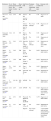
Characteristics of the cases in the literature reporting the use of intravitreal Bevacizumab monotherapy for the treatment of ROP.
| References | No. of patients (eyes) treated with IVB | Mean gestational age (weeks) | Mean birth weight (g) | State before IVB | Treatment used in conjunction with bevacizumab | Dose of IVB used (mg) | Outcome with IVB |
| Travassos and associates, 20079 | 3 (3) | 24.3 | 603 | Anterior NV posterior Zone II (2) | No | 0.75 | Regression of ROP |
| Anterior NV after laser for Zone I (1) | Yes (1 eye salvage) | 0.75 | Regression of ROP | ||||
| Kong and associates, 2008 | 1 (2) | 22 | 350 | Zone I, stage 2+ | No | 0.50 | Regression of ROP |
| Zone II, stage 3+ | No | 0.50 | Regression of ROP | ||||
| Quiroz-Mercado and associates, 200813 | 13 (18) | 29.1 | 1233 | Stage 4A, 4B, 3+Eyes unable to be graded | NoYes (4 eyes salvage) | 1.25 | Regression of ROP (94%)TRD (6%) |
| Mintz-Hittner and associates, 200814 | 11 (22) | 24.3 | 706 | Zone I/Post-Zone II | No | 0.625 | Regression of ROP (100%) |
| AP-ROP | |||||||
| Dorta and associates, 201020 | 7 (12) | 25.5 | 846 | Zone I/Zone II | No | 0.625 | Regression of ROP (100%) |
| APROP | |||||||
| Mintz-Hittner and associates, 201125 (BEAT-ROP study) | 70 (140) | 24.2 | 615 | Zone I stage 3+ | No | 0.625 | Regression of ROP (94%) |
| 24.5 | 689 | Post-Zone II stage 3+ | No | 0.625 | Regression of ROP (95%) | ||
| Axer-Siegel and associates, 201127 | 5 (10) | (24–26) | 620–825 | APROP | No | 0.75 | Regression of ROP (100%) |
| Roohipoor and associates, 201128 | 2 (4) | 28.5 | 1218 | APROP | No | 0.625 | Regression of ROP (100%)2 eyes needed adjuvant laser therapy |
| Harder and associates, 201130 | 12 (23) | 25.1 | 625 | Threshold ROPZone I/Zone IIAPROP | No | 0.375 | Regression of ROP (100%) |
| Wu and associates, 201124 | 23 (41) | 25.7 | 845 | Zone 1/Zone II | No | 0.625 | Regression of ROP (90%) |
| Stage 3+ | Yes (5 eyes salvage) | 4 eyes needed adjuvant laser treatmentVitreus and preretinal hemorrhage (10%) |
The use of IVB as monotherapy for ROP treatment has been reported in a total of 263 eyes of 136 patients from several studies, of which almost all had APROP (Table 4), and the most commonly used dose was again 0.625mg. In 257 (97%) eyes, plus disease disappeared completely and ROP regressed without complication and the anatomical results were favorable. Six eyes needed laser therapy 1.5 month after IVB injection in spite of the initial treatment response. Four (1.5%) eyes experienced vitreous and preretinal hemorrhage, 3 (1%) eyes experienced a progression to tractional RD and one eye (0.37%) developed macular dragging.
The outcome of our series of AP-ROP cases treated with IVB combined with conventional laser therapy is not parallel to the results presented in the literature. Although the effects of bevacizumab to halt the plus disease and anterior neovascularization were remarkable, it does not seem to change the unfavorable outcomes and progression of the severe ROP when used in association with laser photocoagulation or as a salvage therapy in our cases. All of the eyes in our series had Zone 1 or posterior Zone 2 disease with severe stage 3 and stage 4a; therefore the prognosis was poor and the disease regressed totally only in 40% of eyes. However it is difficult to make any conclusion with this information since the timing of therapy and case selection is still a problem in APROP cases. Our impression is that, the severity of stage 3 disease plays an important role in response to IVB therapy. When it is a severe stage 3 disease where extraretinal dilated abnormal vessels in ridge are highly elevated through vitreous, IVB therapy leads to conversion of these vessels to a fibrotic tissue causing traction of the retina. However when it is a mild stage 3 disease in Zone 1 with a flat neovascularization, those eyes respond well to IVB therapy.
Another cause of poor outcome of the IVB therapy in our series is its use as a salvage therapy which occurred in most (83%) of the eyes. Which means that, IVB was given to those eyes that did not respond well to the laser treatment and had tendency to progress to stage 4. The high rate of anterior segment ischemia (15.3%) in our cases added to the poor outcome. This is possibly because of the need for huge amount of laser photocoagulation in very large avascular areas in Zone 1 disease. The mean number of burns in our cases that developed anterior segment ischemia was 2452±311 spots. Anterior segment ischemia has been reported by Fallaha and associates, where a 2.3% rate of anterior segment ischemia was seen in their cohort of 91 eyes.35 Kaiser and Trese reported on 8 eyes of 5 patients referred from other clinics who developed cataracts, iris atrophy and hypotony, all symptoms indicative of anterior segment ischemia following laser photoablation.36 Lambert et al. reported a series of 10 eyes that developed cataracts and phthisis bulbi secondary to anterior segment ischemia following laser photoablation for threshold ROP where the mean number of burns applied was 2532±856 similar to our cases.37
In conclusion, despite the limitations of this study (its retrospective nature, absence of a control group and a small number of patients and limited functional results) the addition of IVB after the laser ablation slightly decreased the progression rate in severe AP-ROP eyes. Prompt regression of iris neovascularization encourages its use in cases with pupillary rigidity to allow for laser treatment. When used as a salvage therapy it may not change the overall result dramatically. Limited literature on this subject indicates that prospective randomized controlled trials are warranted.
Conflicts of interestThe authors have no conflicts of interest to declare.


