
To investigate the effect of a + 2.00 D fogging lens on accommodative behavior during measurements with an open-view handheld wavefront autorefractor, considering refractive status and fogging duration.
MethodsParticipants underwent six consecutive 10-second measurements per eye using an open-view autorefractor: three without and three with a fogging lens. Subjects were classified as myopic, emmetropic or hyperopic based on the mean spherical refraction of the right eye. Bland–Altman analysis assessed device repeatability and the effect of fogging (with vs without), both overall and by refractive group. The influence of acquisition time was evaluated by comparing the first 5 s with the full 10-second recordings.
ResultsSixty-one young adults (25.02 ± 6.16 years; spherical refraction -0.80 ± 2.21 D) were included: 34.4% myopes, 42.6% emmetropes and 22.9% hyperopes. Ten-second measurements showed excellent repeatability, with mean differences near zero and 95% limits of agreement within ±0.50 D, regardless of fogging. Shortening acquisition to 5 s maintained negligible bias but increased variability by ∼20%. The fogging lens induced heterogeneous responses across refractive groups. In myopic and emmetropic eyes, most showed no change (52% and 58%), and accommodative relaxation occurred infrequently (10% and 17%). In contrast, 43% of hyperopic eyes showed accommodative relaxation, with a mean refractive shift of 0.84 D, suggesting a clinically meaningful response.
ConclusionThe effect of a + 2.00 D fogging lens depends on refractive status. Routine use may provide limited additional benefits in myopic and emmetropic subjects, whereas hyperopic individuals appear more likely to benefit from its selective use.
Autorefraction is a cornerstone of optometric and ophthalmologic practice, providing an objective assessment of refractive error. A key limitation, however, especially in children and young adults, is its vulnerability to inaccuracies induced by accommodation. This physiological process allows the eye to change the shape of the crystalline lens to focus on objects at varying distances and is mediated by the ciliary muscles under neural control. When active during autorefraction, accommodation can produce an artificial myopic shift.1 This problem is particularly evident with closed-view autorefractors, which present subjects with an internal fixation target located only a few centimeters inside the instrument. The artificial nature of the target and the enclosed viewing environment tend to stimulate, rather than relax, accommodation2 (instrument-induced myopia). To mitigate this effect, and the resulting over-minusing of spherical refraction, closed-view devices employ blurred fixation targets, dim illumination, or fogging lenses within the optical path. While these strategies reduce accommodative demand, they are only partially effective and frequently insufficient to eliminate the need for cycloplegia in some subjects.3-5
To address these limitations, open-view autorefractors have been developed to allow subjects to look through the device and fixate on a distant external target, thereby better mimicking natural viewing conditions. This open-view design promotes a more relaxed accommodative state compared to closed-view systems. Studies have shown that open-view systems yield measurements closer to subjective refraction and to those obtained under cycloplegia, particularly in non-presbyopic individuals.6-8 However, even with this design, residual accommodation may persist in subjects with high accommodative tone, most notably children, young adults and individuals with latent or uncorrected hyperopia, potentially requiring additional cycloplegic measurements for an accurate assessment. To further control accommodation, fogging lenses are often used in open-view systems. Fogging involves introducing a low-powered plus lens (typically between +1.00 D and +2.00 D)9–11 in the viewing path to blur the image on the retina, thereby encouraging the eye to relax accommodation. This technique has been shown to reduce the accommodative response and improve the agreement between objective and cycloplegic refraction.9,10,12 Studies using open-view wavefront aberrometry based autorefractors have confirmed that fogging with low-powered plus lenses can induce measurable reductions in accommodative response within seconds. This response is dose- and time-dependent wherein mild fogging for short durations (e.g., +1.00 D for 5 - 30 s) can provide significant accommodation relaxation, whereas excessive fogging (+3.00 D or more) or long application durations may result in paradoxical re-accommodation due to the eye reverting to its resting or “dark focus” state.1,13,14
This study aims to evaluate the impact of fogging lenses on accommodative behavior when using an open-view handheld wavefront autorefractor, the QuickSee Free Pro Keratometry (QSF Pro; PlenOptika Inc., USA), which allows fogging lenses to be magnetically attached to the viewing path. In addition, we assess how the effect of fogging varies according to the subject’s refractive status. Previous studies have shown that myopes exhibit minimal changes due to their lower accommodative demand, whereas emmetropes, and particularly hyperopes, demonstrate stronger accommodative responses. Notably, hyperopes appear to benefit the most from fogging, underscoring the importance of tailoring fogging strategies to refractive status, especially in non-cycloplegic conditions.10 Finally, we evaluated the influence of fogging lens application time by comparing measurements obtained without fogging to those acquired with a shortened acquisition of 5-seconds versus the device default setting of 10-second application.
Material and methodsQuickSee free pro wavefront autorefractorThe QSF Pro is a handheld open-view wavefront autorefractor that employs a simplified Shack-Hartmann optical architecture. It measures refractive error (including high-order aberrations) sequentially in each eye, with a spherical range of −13.00 to +10.00 D and cylindrical ranges up to 8.00 D Additional details on the device’s technical performance and proprietary data processing algorithms have been reported elsewhere.15,16 The firmware of the device used in the study was the most recent version provided by the manufacturer (v6.0.3, released May-2025).
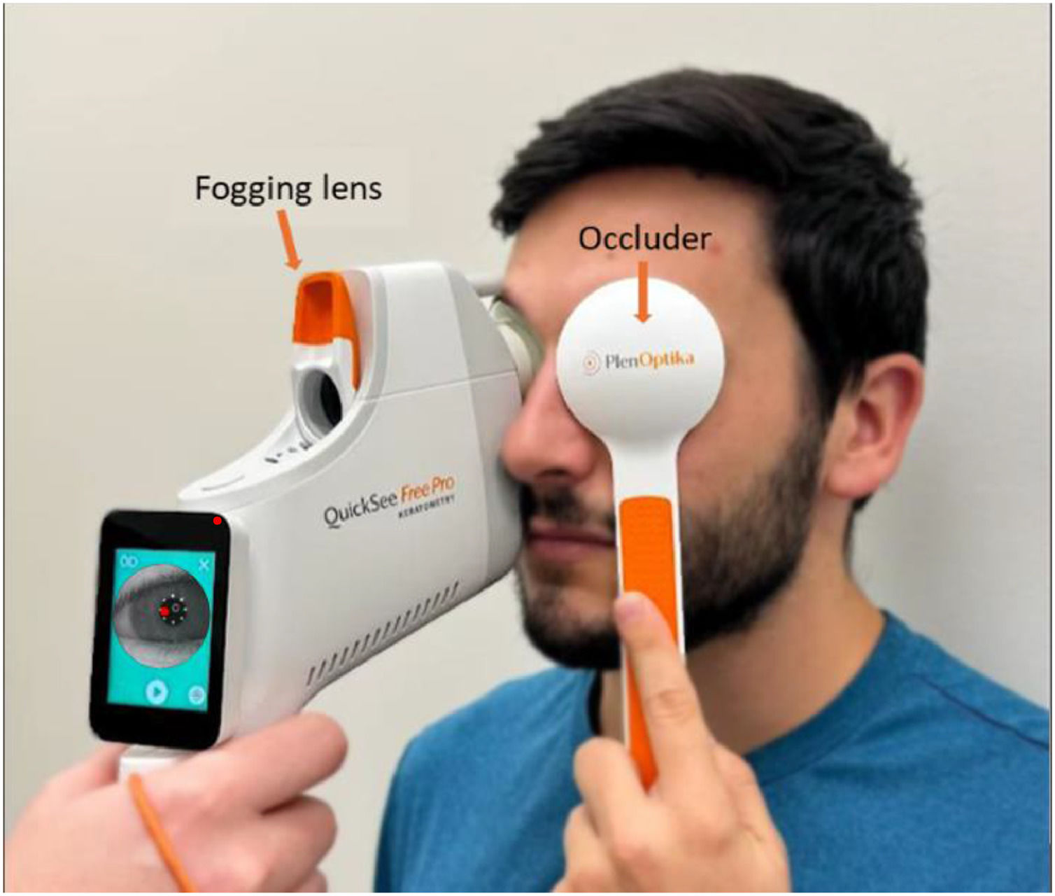
During measurement, the operator aligns the viewport with the subject’s eye using a horizontal guideline printed on the side of the device. The position is stabilized over a 5- or 10-second measurement by gently bracing the device against the subject’s face at two contact points. Crucially, the viewport features a completely open and transparent optical channel, providing an unobstructed central field of view. This design ensures that during the measurement subjects fixate on a distant external target under natural viewing conditions, minimizing proximal accommodation (instrumental myopia) that typically occurs with closed-field systems. Removable fogging lenses are magnetically attached to the outer viewport in the subject’s line of sight to facilitate accommodative relaxation during measurement (Fig. 1) without affecting the wavefront aberration signal detected by the measurement optics of the device.
Research protocol
Participants were recruited from among students and staff at the School of Engineering of the Autonomous University of Madrid. Inclusion criteria were: (1) no history of ocular surgery or eye disease, (2) age between 18 and 39 years, (3) corrected visual acuity (VA) of 0.1 LogMAR or better in each eye, (4) no use of systemic or ocular medications known to affect vision, and (5) spherical refractive error within −13.00 D to +10.00 D and astigmatism of ≤ 8 diopters. The research protocol was approved by the University’s Research Ethics Committee and conducted in accordance with the tenets of the Declaration of Helsinki. All participants provided written informed consent after receiving both verbal and written explanations of the study procedures.
Each participant completed six 10-second measurements per eye using the portable autorefractor. The first three measurements were taken without a fogging lens, followed by three with a + 2.00 D fogging lens mounted on the device. For the latter, total exposure to fogging included both the 10-second acquisition period and the brief alignment time required for the operator to position the device on the eye (typically <3 s). During all measurements, participants were instructed to fixate on a visual acuity chart positioned at 6 m, while the contralateral eye was occluded with the handheld occluder supplied with the device to help promote pupil dilation (Fig. 1). Between measurements, a rest period of 30–60 s was allowed with no visual correction. To evaluate the impact of fogging duration, dynamic recordings were analyzed using either the first 5 s or the full 10-second interval of each measurement.
Data analysisSubjects were categorized into three refractive groups: myopes, hyperopes, and emmetropes. Classification was based on the average spherical component derived from the three right-eye refraction measurements obtained under each condition (with and without the fogging lens). To determine refractive status, the more positive of the two averaged values was selected, and the following criteria were applied: ≥ +0.50 D for hyperopia, ≤ −0.50 D for myopia, and between −0.50 D and +0.50 D for emmetropia, consistent with commonly used clinical cut-offs in refractive error classification.17
Dynamic recordings were acquired at a sampling frequency of 8 frames per second. Each measurement lasted 10 s and consisted of two consecutive 5-second segments. For analysis, the refractive value for each segment corresponded to the temporal mean of all frames recorded within that interval. The 10-second value therefore represented the average of the two 5-second segment means. All repeatability and comparative analyses were based on these temporally averaged values rather than on single time-point measurements.
Device repeatability was assessed using Bland-Altman analysis: for each subject and condition (with and without fogging), pairwise differences among the three measurements (M1-M2, M1-M3, M2-M3) were calculated to construct Bland-Altman plots. From these, the mean difference (bias) and 95% limits of agreement (LOA), calculated as ±1.96 times the standard deviation of the differences, were derived as repeatability metrics. The bias is a measurement of accuracy of the device (assuming that the true refraction value is the average of M1, M2, and M3). The standard deviation and the 95% LOA provide information about the variability of the device, in terms of how close repeated measurements are to each other, regardless of whether they are close to the true value. Analyses were performed for the overall study population and stratified by refractive group, under both fogging and non-fogging conditions. Repeatability under fogging was also examined across different application times (5 and 10 s). In all cases, results are provided for the spherical component (S) of refraction as well as for spherical equivalent (M) and vectorial components of the astigmatism (J0 and J45) .18
The effect of the fogging lens within each refractive group was further examined using Bland-Altman analysis, comparing the average of the three refraction measurements obtained with and without the lens across all application times. Differences in spherical refraction were categorized as follows: no effect (NE) when the difference was between −0.125 D and +0.125 D, accommodation induction (AI) when difference was ≥ +0.125 D, and accommodation relaxation (AR) when difference was ≤ −0.125 D For each refractive group and for the overall sample, both the proportion of participants and the mean spherical difference produced by the fogging lens were calculated within the NE, AR, and AI categories to quantify the direction and magnitude of its accommodative influence. Additionally, to directly quantify the statistical effect of the +2.00 D fogging lens, paired t-tests were performed comparing measurements with and without the lens for the overall population and separately for each refractive group (myopes, emmetropes, and hyperopes). A p-value < 0.05 was considered statistically significant.
ResultsStudy populationA total of 61 participants were included in the study. The mean age of the cohort was 25.02 ± 6.16 years (mean ± standard deviation), with a range of 18 to 39 years. The sample included 30 males (49.2%) and 31 females (50.8%). According to the refractive classification previously described, 21 participants (34.4%) were classified as myopic, 26 (42.6%) as emmetropic, and 14 (22.9%) as hyperopic. Participants were recruited from students and staff of the Universidad Autónoma de Madrid to obtain a cohort spanning a range of refractive conditions. Therefore, the refractive distribution of the sample was not intended to represent the prevalence of refractive errors within the university population.
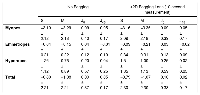
Descriptive characteristics of the study population, including the mean refractive values for each group with and without the application of fogging lenses (10-second measurements), are summarized in Table 1. Refractive outcomes are reported as spherical refraction (S) and in vector notation (M, J0, J45) derived from Fourier analysis, following the standard convention used in refractive error studies.18
Average refraction values by subject group for 10 s measurement. Data is presented as mean ± standard deviation (expressed in diopters) for the spherical refraction (S) and three vector components (M, J0, J45) across refractive groups, measured without (No Fogging) and with a + 2.00 D fogging lens.
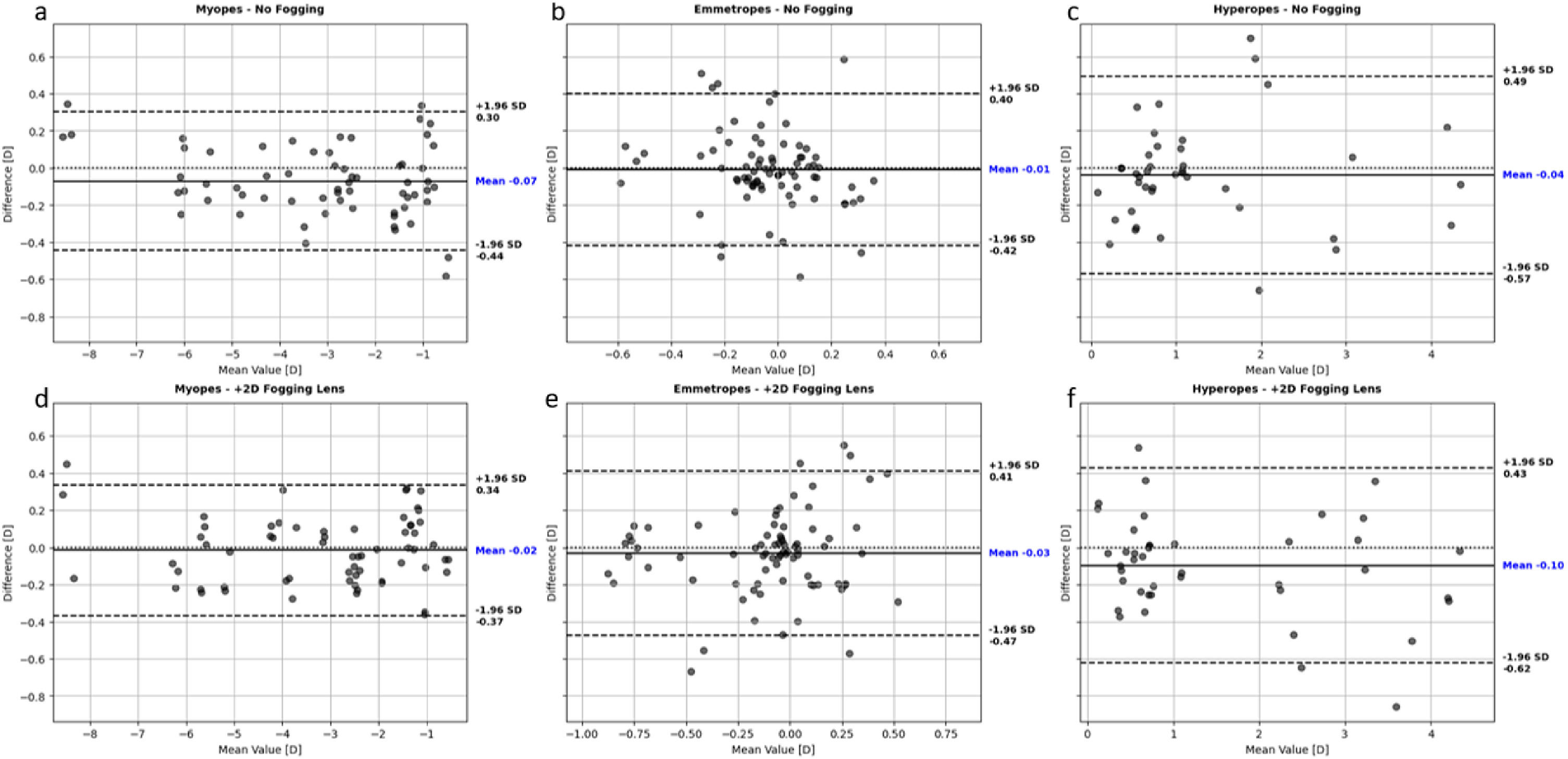
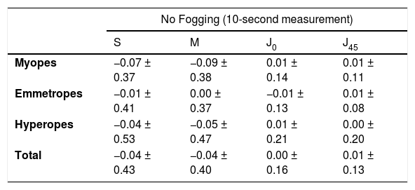
Fig. 2 shows Bland-Altman plots of spherical differences in diopters across repeated 10-second measurements for each refractive group, with (bottom row) and without (top row) fogging lenses. Table 2 summarizes detailed results for all refractive components (S, M, J0 and J45), reported by refractive group and for the overall population, including both 10- and 5-second fogging application times.

Repeatability of spherical refraction (in diopters) across refractive groups, with and without fogging lens application (10-second measurement). The top row (a-c) shows Bland-Altman plots without the fogging lens, while the bottom row (d-f) shows results with the lens. Columns correspond to refractive groups: myopes (first), emmetropes (second), and hyperopes (third).
Repeatability of the QuickSee Free Pro with human eyes in diopters, detailed results. Data are provided as Bias ± 95% LOA (in diopters) of the corresponding Bland Altman Analysis. As a reference, Bland-Altman plots for 10-second measurements with and without fogging lens can be seen in Fig. 2.
Without fogging, all groups showed low variability in the spherical (S) and vector components (M, J0, J45), with mean differences close to zero, indicating high measurement consistency. With the +2.00 D fogging lens applied for 10 s, repeatability remained high in myopes, while emmetropes and hyperopes showed slightly greater variability in the spherical component, suggesting that fogging has a marginally bigger effect in these groups. For the 5-second fogging application, bias remained constant and close to zero, but variability increased across all groups, indicating, as expected, that shorter acquisition times lead to slightly worse repeatability (increase of around 20% in LOA). In contrast, the bias and LOA of J0 and J45 components remained near zero and almost constant in all cases, indicating that cylindrical measurements were not affected by accommodation response under all conditions.
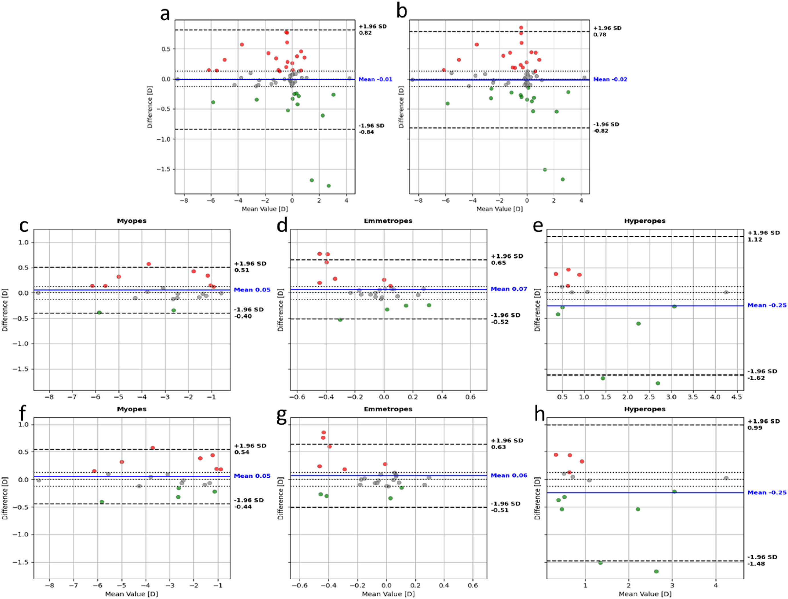
Effect of fogging lensesAverage differences in spherical refraction with and without the fogging lens (Bland-Altman bias; Fig. 3) were negligible when the overall population was analyzed, with variability (standard deviation and 95% LOA) only slightly greater than that observed in the repeatability tests. For the 10-second fogging-lens application, repeatability results were −0.04 ± 0.43 D (Table 2), compared with a fogging-no fogging related difference of −0.01 ± 0.83 D (paired t-test, p = 0.61; Fig. 3, left). Similarly, for the 5-second application, repeatability results were −0.04 ± 0.64 D (Table 2), while the fogging-related difference was −0.02 ± 0.80 D (p = 0.71; Fig. 3, right). Overall and considering the whole study population, reducing the fogging-lens application time from 10 to 5 s did not appear to produce meaningful changes in either bias or measurement variability (Fig. 3, Table 3).

Bland–Altman plots showing differences in spherical refraction between measurements with and without fogging lenses for 10-second (a) and 5-second (b) acquisitions in the overall sample, and by refractive group for 10-second (c-e) and 5-second (f-h) measurements (myopes, emmetropes and hyperopes, respectively). Dotted lines indicate zero difference and the limits of the neutral effect (−0.125 D to +0.125 D). Red points (above +0.125 D) represent accommodation induction (AI), green points (below −0.125 D) indicate accommodation relaxation (AR), and grey points denote no effect (NE).
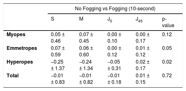
Effect of +2D fogging lens in human eyes, detailed results. Data are provided as bias ± 95% LOA of the corresponding Bland-Altman Analysis (Fig. 3 and 4) for the spherical and vectorial components of the refraction. All results included in the table are in diopters.
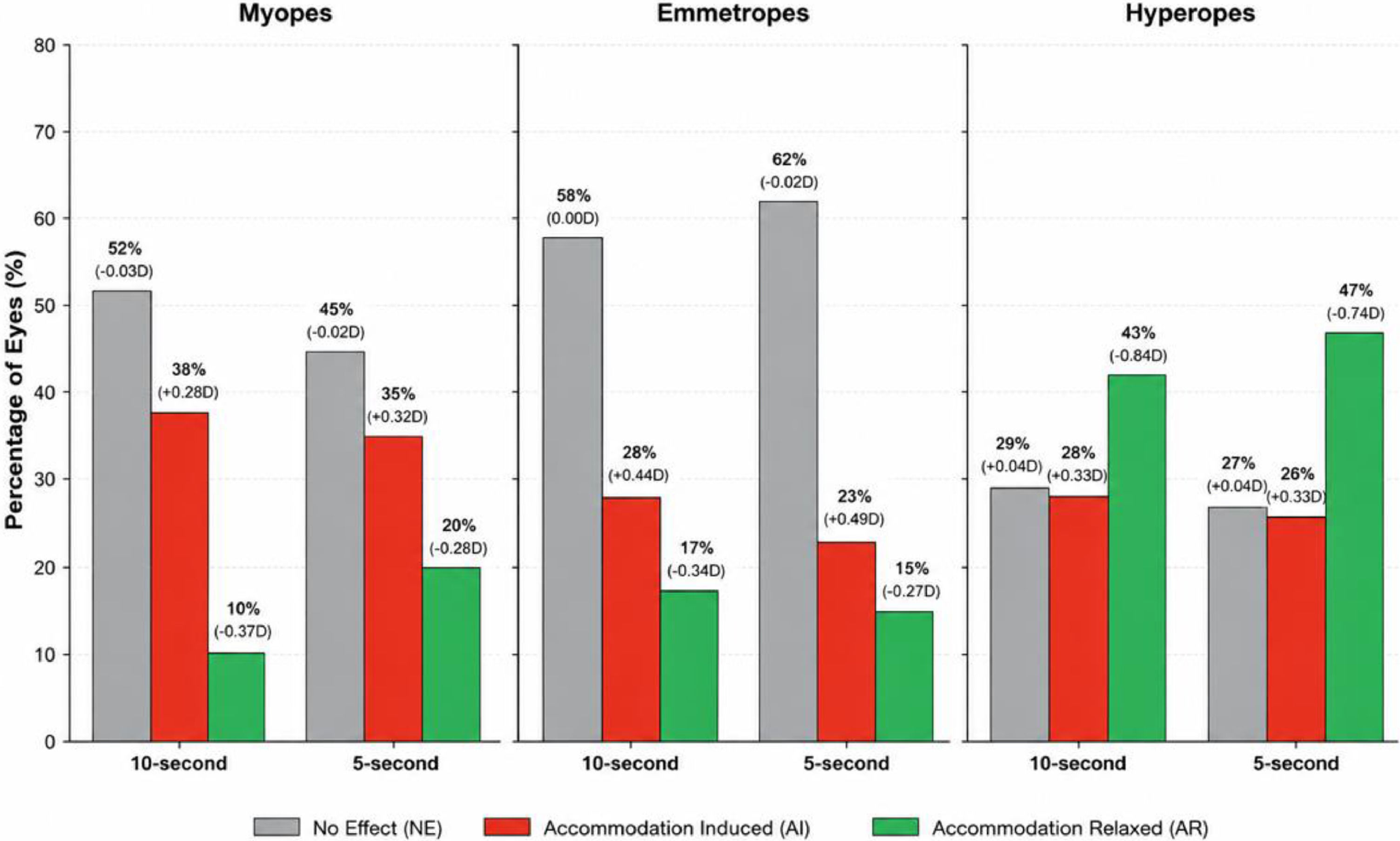
In contrast, when the same data were analyzed by refractive group, larger bias values emerged in all categories, and the 95% LOAs were consistently wider than those from repeatability analysis. To better illustrate these findings, central and bottom rows from Fig. 3 present the same analysis as the top row but stratified by refractive group, and Table 3 summarizes the numerical results, including changes across all vector components of refraction for each group and for the overall sample. To further assess the effect of the fogging lens within each refractive group, Fig. 4 summarizes the proportion of participants exhibiting no effect (NE), accommodation induction (AI), or accommodation relaxation (AR), together with the corresponding mean change in spherical refraction for each response category. Results are presented separately for the 10- and 5-second application times, allowing direct comparison of the accommodative response distributions across refractive groups and measurement durations.

Distribution of accommodative responses following application of the +2.00 D fogging lens. The percentage of eyes exhibiting no effect (NE), accommodation induction (AI), or accommodation relaxation (AR) is shown for myopic, emmetropic, and hyperopic subjects after 10- and 5-second applications. Values above each bar indicate the proportion of eyes in each category, while values in parentheses correspond to the mean spherical refractive change (D) for that subgroup.
When assessed using Bland-Altman analysis, comparisons between the 10- and 5-second fogging lens application times (Fig. 3, central and bottom rows) showed no meaningful differences in either bias or 95% limits of agreement for any refractive group, suggesting that both application times yield comparable results at the population level. However, this global analysis masks differences that become apparent when the effect of the fogging lens is examined within refractive groups and response categories (no effect, accommodation induction, or accommodation relaxation; Fig. 4). From this perspective, the 5-second application produced results more consistently aligned with the intended purpose of the fogging lens, namely, promoting accommodation relaxation. In the overall population, reducing the application time from 10 to 5 s increased the proportion of eyes exhibiting accommodation relaxation (from 20% to 25%), decreased the proportion showing induced accommodation (from 31% to 28%), and resulted in only a minor change in the proportion of eyes showing no measurable effect (49%vs. 47%). Importantly, this trend was also observed when the analysis was performed separately within each refractive group, rather than when only population-level Bland-Altman metrics were considered.
Finally, to facilitate the clinical interpretation of the response-based subgroup findings, Fig. 5 compares the accommodative changes induced by the +2.00 D fogging lens with the intrinsic repeatability of the device, represented by the 95% limits of agreement (LOA) obtained from repeated measurements.

Comparison between the accommodative changes induced by the +2.00 D fogging lens and the intrinsic repeatability of the device, represented by the 95% limits of agreement (LOA) obtained from repeated measurements. Mean spherical refraction changes for each accommodative response category are shown relative to the repeatability limits of the instrument for myopic, emmetropic and hyperopic subjects.
In myopic eyes, the fogging lens relaxed accommodation in 20% of subjects. The average relaxation among these subjects was 0.22 D, which remained within the device's intrinsic repeatability limits (95% LOA), Fig. 5. Overall, the lens produced no measurable effect in the majority of myopic subjects (45%) and induced accommodation in the 35% of cases, with an average myopic shift of about 0.3 D, also remaining within the device’s intrinsic repeatability for this subgroup (Fig.5).
A similar pattern was observed in emmetropic eyes. The fogging lens had no measurable effect in most subjects (62%). Accommodation relaxation occurred in only 15% of cases, with an average magnitude smaller than the device’s intrinsic repeatability (Fig. 5). In contrast, accommodation was induced in 23% of emmetropic eyes, with average changes in spherical refraction (0.49 D) slightly exceeding the expected intrinsic repeatability for this subset (Fig. 5).
Conversely, hyperopic subjects exhibited a markedly different response. Although the average relaxation across all hyperopic eyes was modest (0.25 D; Table 3, 5-second comparison), Figs. 4,5 show that accommodation was relaxed in 47% of eyes with an average magnitude of 0.74 D, clearly exceeding the intrinsic repeatability observed in repeatability testing for this group (95% LOA, Fig. 5). Accommodation induction was observed in 26% of hyperopic eyes, with an average change of 0.33 D, which was within the expected intrinsic repeatability determined for this group (Fig. 5).
DiscussionThis work investigated the influence of a + 2.00 D fogging lens on refractive measurements obtained with a portable open-view wavefront autorefractor, assessing its effect across different refractive groups and application times.
Our results indicate that the effect of the +2.00 D fogging lens differs according to refractive status. In myopic and emmetropic subjects, accommodative relaxation was observed in only a small proportion of participants, and the magnitude of the relaxation was generally comparable to or smaller than the device's intrinsic repeatability limits (95% LOA). Conversely, many subjects exhibited either no measurable effect or accommodative induction, suggesting that the clinical benefit of routine fogging may be limited in these groups. Statistical comparison of spherical refraction with and without the fogging lens showed no significant differences in myopes (mean diff = 0.05 D, p = 0.12) or emmetropes (mean diff = 0.07 D, p = 0.05), whereas hyperopes exhibited a small but statistically significant difference (mean diff = −0.25 D, p = 0.02 for the 10-second application; mean diff = −0.25 D, p = 0.01 for the 5-second application), indicating that the lens produced its greatest measurable effect in this refractive group. Accordingly, our findings suggest that routine application of the fogging lens in myopic and emmetropic subjects is unlikely to provide substantial additional benefit and, in some individuals, may introduce additional measurement variability (Figs. 4 and 5).
In contrast, hyperopic subjects exhibited the highest proportion of accommodative relaxation, with 43% of eyes showing relaxation and an average spherical refractive change of approximately 0.75 D, well above the device's intrinsic repeatability limits (95% LOA). These findings suggest that hyperopic individuals are more likely to benefit from the use of fogging lenses. Regarding application time, the distributions of accommodative responses observed after 5 and 10 s were broadly comparable. Although the 5-second protocol showed a slightly higher proportion of accommodation relaxation and a lower proportion of accommodation induction, these differences were modest, and no significant differences were observed in the overall population-level bias between the two application times (overall mean diff = −0.01 D, p = 0.72 for 10 s; mean diff = −0.02 D, p = 0.61 for 5 s). Together, these findings suggest that extending the fogging period beyond 5 s provides limited additional clinical benefit under the conditions evaluated in this study.”
Our findings are partially consistent with previous reports on the effect of fogging lenses in open-field autorefractors. Queirós et al.10 evaluated the impact of a + 2.00 D fogging lens in a cohort of 142 young adults (22.3 ± 3.7 years) using a well-established binocular open-view desktop system (WAM-5500, Grand Seiko Co. Ltd). In their study, the average accommodation relaxation induced by the lens was comparable to that achieved under cycloplegia in terms of spherical equivalent refraction. Notably, they analyzed these differences by refractive group, reporting the spherical equivalent (M) of average accommodation relaxation of 0.4 D for myopes (n = 38), 0.3 D for emmetropes (n = 42), and 0.28 D for hyperopes (n = 62). These results contrast with our findings, where we observed mean differences of only 0.06 D for myopes (n = 21), 0.07 D for emmetropes (n = 26), and 0.24 D for hyperopes (n = 14). A plausible explanation for this discrepancy is the difference in viewing conditions between the two studies. In the present study, measurements were intentionally performed under monocular viewing conditions, consistent with the intended clinical use of the QuickSee Free Pro, with the contralateral eye occluded. This eliminates the influence of convergence and may reduce both the amplitude and stability of accommodation, as the accommodative-convergence mechanism is not fully engaged.19 In contrast, Queirós et al.10 performed binocular measurements, preserving the natural coupling between accommodation and convergence, which typically yields more stable and physiologically representative accommodative responses. Therefore, differences in viewing conditions should be considered when comparing the results of both studies. Nevertheless, viewing conditions alone are unlikely to fully explain the discrepancies observed, particularly in the myopic subgroup. As suggested by Queirós et al. and other authors,20,21 accommodative relaxation in myopic participants may be influenced by changes in pupil diameter under fogging conditions, as larger pupils can increase optical aberrations and affect the refractive endpoint measured by the autorefractor. Although the QSF Pro acquires dynamic wavefront data, including pupil diameter and higher-order aberrations such as spherical aberration, these parameters were beyond the scope of the present study and were therefore not analyzed. Future studies incorporating these measurements could help clarify the optical mechanisms underlying the inter-subject variability observed during fogging. Finally, differences in the duration of fogging lens application and the variability observed within each refractive group were not reported in the Queirós study, limiting a more precise comparison with our findings.
Bui et al. investigated the effect of a + 2.00 D fogging lens in 53 children aged 4 to 12 years with varying degrees of hyperopia.22 In that study, the authors employed an earlier model of a portable open-view autorefractor (QuickSee, PlenOptika Inc, USA),23,24 which relied on wavefront sensing technology similar to that of the monocular QSF Pro used in the present work but differed in several important design aspects. These design differences included a binocular measurement configuration, a reduced field of view, and the placement of the fogging lens within the measurement path and requiring post hoc subtraction of its optical power from the refractive results. In contrast to the findings of the present study and those reported by Queirós et al., Bui et al. found no statistically significant difference in spherical refraction with and without the fogging lens (mean difference = −0.085 D; p = 0.51), which they primarily attributed to effective accommodative control provided by a distant, non-accommodative, colour-changing fixation target. Beyond these considerations, discrepancies between studies may also reflect differences in subject populations and fixation behaviour (children versus adults), as well as methodological factors such as monocular versus binocular measurements and the use of static versus dynamic fixation stimuli (visual acuity chart in the present study versus colour-changing lights in Bui et al.).
In another similar study, Turull-Mallofré et al.13 also examined the influence of fogging lens power and application duration on accommodation in young adults using a wavefront aberrometer and open-view measurements. They found that lower-power lenses (+1.00 D and +2.00 D) produced the greatest accommodation relaxation, with 59.3% and 46.9% of participants, respectively, showing a median relaxation ≥ 0.125 D Regarding duration of fogging lens application, no consistent trend was observed for all lens powers; however, maximum relaxation with all lenses generally occurred within the first ≈5 s of measurement, which is consistent with our results. Furthermore, the authors also reported substantial inter-individual variability with all lenses, which increased for longer fogging lens application durations. They suggested that this variability, also seen in our study (Table 3), might stem from the wide inter-subject differences in stimulus–response function and tonic accommodation previously reported in the literature.25 Unfortunately, since the results were not stratified by refractive group, it is unclear whether refractive status contributed to the observed variability. Furthermore, although some findings were consistent with ours, methodological differences, particularly their use of a desktop prototype aberrometer where one eye viewed the target through the fogging lens while the contralateral eye was measured in a closed-view configuration, limit direct comparison with our results and with other studies using fully open-view monocular or binocular systems.
Finally, several limitations of the present study should also be acknowledged. First, measurements were always performed in the same sequence, with the non-fogging condition preceding the fogging condition. Although this protocol was selected to reflect the intended clinical workflow, a randomized crossover design would have further minimized the possibility of measurement-order effects. However, the refractive-group specific response observed, with significant effects primarily in hyperopic subjects and minimal changes in myopic and emmetropic eyes, suggests that the findings cannot be explained solely by a measurement-order effect. Nevertheless, future randomized crossover studies would be valuable to further confirm these findings. Second, participants were classified based on non-cycloplegic objective refraction, which may not fully represent true refractive status, particularly in young or hyperopic subjects with active accommodation. Although the more positive average of repeated measurements was used to reduce the influence of residual accommodation, some misclassification cannot be excluded. In particular, latent hyperopic subjects may have been classified as emmetropic, reducing the separation between these groups and potentially attenuating, rather than exaggerating, the apparent effect of the fogging lens. A classification based on cycloplegic or subjective refraction would therefore improve group separation and strengthen the interpretation of accommodative responses. Furthermore, the relatively small hyperopic subgroup also limited the statistical precision of the estimated effect size and reduced the ability to detect more subtle accommodative responses or variability patterns. Although the findings in this subgroup were internally consistent across the different analyses performed, they should be interpreted with appropriate caution until confirmed in larger cohorts. Future studies should therefore include larger and more balanced samples and incorporate cycloplegic measurements alongside fogging protocols to better validate accommodative relaxation and individual control. Additionally, assessing accommodation response under different fogging conditions such as monocular, binocular, or applied to the contralateral eye, would allow direct comparison of measurement paradigms and help clarify the influence of convergence and device design. Such work would further contextualize the present findings within the broader literature.
ConclusionThis study demonstrates that the effect of a + 2.00 D fogging lens used with the open-view wavefront autorefractor is strongly dependent on refractive status. In myopic and emmetropic subjects, the lens did not reliably induce accommodative relaxation. Most eyes showed no measurable change, and a substantial proportion exhibited accommodation induction, with mean differences small and not statistically significant. These findings suggest that routine use of the fogging lens in these groups is unlikely to provide substantial additional benefit and may potentially increase measurement variability.
By contrast, hyperopic subjects exhibited larger mean differences and a substantial proportion of eyes showing accommodative relaxation beyond the device's intrinsic repeatability, suggesting that this population is more likely to benefit from the use of a + 2.00 D fogging lens. Moreover, similar accommodative response distributions were observed after both 5- and 10-second applications, indicating that extending the fogging period beyond 5 s may offer little additional practical benefit. Overall, these findings support a more selective use of fogging lenses in monocular open-view autorefractors according to the subject's refractive status, rather than their routine application in all patients.
Author contributions and consent to publishP.B performed the measurements and analyzed the data presented in the figures and tables. G.G., P.E., A.H. and M.R. designed, implemented, and supervised the device software. A.W., I.C. and A.L. built the device and supervised its proper functionality. J.O., F.F., E.L., S.D. and D.L. prepared and supervised the regulatory documentation and clinical trial protocol. P.B., E.L. and J.O. wrote the main manuscript text. All authors reviewed the manuscript. All authors listed on the title page consent to the publication of the text, data and images contained within this paper.
FundingResearch related to the autorefractor technology used in this work was partially supported by the Spanish Ministry of Science and Innovation (TED2021-131541A-I00), the US National Eye Institute of the National Institutes of Health (R44EY025452), and Translational Research Institute for Space Health (NASA NNX16AO69A, USA). The content is solely the responsibility of the authors and does not necessarily represent the official views of the National Institutes of Health or the Spanish Ministry of Science and Innovation.
Ethics approval and consent to participateThe study was approved by the Autonomous University of Madrid Research Ethics Committee. The study adhered to the tenets of the Helsinki Declaration at all stages. We obtained written informed consent from all participants.
Eduardo Lage, Shivang R. Dave, Daryl Lim, Ignacio Casares, Paula Egido, Alfonso de Lara, Flavia Fondevila and Alec Wehse are stock owners or have stock options of PlenOptika Inc. Eduardo Lage, Shivang R. Dave and Daryl Lim are authors in patents filled by PlenOptika Inc. Pablo Bargalló, Ignacio Casares, Gonzalo Guerrero, Paula Egido, Flavia Fondevila, Javier Ortega-Julia, Alec Wehse, Alberto Hernandez, Miguel Rangel and Alfonso de Lara are or have been employees of PlenOptika Inc. Shivang R. Dave and Daryl Lim and Eduardo Lage have executive roles in PlenOptika Inc. The study described in this work was partially funded by PlenOptika Inc; other sources of funding related to this work are disclosed in the Funding Information section of the manuscript.
www.plenoptika.com